Description of the structure of the amphibian brain. Amphibian Anatomy: An Overview
, reptiles (reptiles), birds, their nests, eggs and voices, and mammals (animals) and traces of their vital activity,
20
colored laminated definition tables, including: aquatic invertebrates, diurnal butterflies, fish, amphibians and reptiles, wintering birds, migratory birds, mammals and their tracks,
4
pocket field determinant, including: inhabitants of reservoirs, birds of the middle zone and animals and their traces, as well as
65
methodological benefits And 40
educational and methodological films By methods carrying out research work in nature (in the field).
Amphibian Anatomy: An Overview
Body structure or anatomy.
Body divided into head, torso, tail (only in caudates and legless animals) and limbs, which may be absent. Head mobile, connected to the body. The skeleton and spine are divided into departments. Ribs, if any, are attached to the trunk vertebrae.
Amphibians have two pairs of primary five-toed fingers limbs; the rudimentary forms of the neck provide them with the ability to move their heads independently.

Leather naked, devoid of scales. Epidermis rich in multicellular glands, which ensure the presence of a liquid film on the surface of the skin, without which gas exchange is impossible during skin respiration. The epidermis is multilayered, the corium is thin, but richly saturated with capillaries.
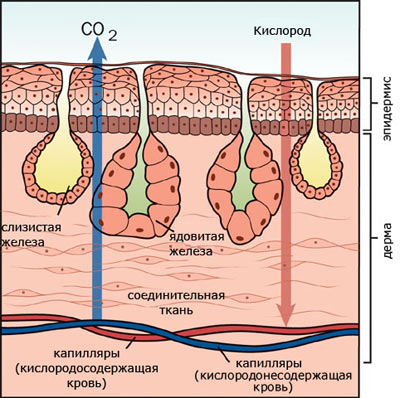
In the lower layers of the epidermis and in the corium are located pigment cells, causing species-specific coloring.
Limb skeleton formed by the skeleton of the limb girdle and the skeleton of the free limbs.
Shoulder girdle lies in the thickness of the muscles and includes paired shoulder blades, collarbones and crow bones connected to the sternum. Skeleton forelimb consists of the shoulder (humerus), forearm (radius and ulna bones) and hand (bones of the wrist, metacarpus and phalanges of the fingers).
Pelvic girdle consists of paired iliac ischial and pubic bones fused together. It is attached to the sacral vertebra through the ilia. Included in the skeleton hind limb includes the thigh, lower leg (tibia and fibula) and foot. Bones of the tarsus, metatarsus and phalanges of the fingers. The sacrum consists of only one vertebra.
Propulsion system.
The movement pattern of amphibians is quite monotonous and can be reduced to two main types.
Fossil and modern tailed amphibians have retained their characteristic fish the main type of movement is with the help of strong lateral bends of the whole body, but with support on short legs when moving on the ground. With short limbs, the lateral bends of the body increase the length of the step, and the bends of the tail help maintain balance. When moving in water, the limbs do not play any noticeable role. Legless animals also move using the bends of the entire body.
Tailless amphibians move on land jumping, lifting the body into the air with a sharp push of both hind limbs. Short-legged species, such as toads, in addition to jumping, can slowly step, sequentially rearranging limbs.
Tailless in the water swim, vigorously working with the hind limbs (breaststroke style, but without the participation of the front limbs). It is believed that powerful hind limbs developed as an adaptation to swimming, and only later were used for jumping on land.
Amphibians have a rather large, wide head, which goes directly into wide and short body. The frontal and parietal bones are fused into the paired frontoparietal bone. IN skull it is characteristic that the maxillopalatine apparatus and the quadrate bone are motionlessly connected to the skull; the two condyles of the skull belong to the first cervical vertebra that actually merged with it, so that the first vertebra of amphibians is essentially the second.
Brain amphibians differ from the fish brain in the greater development of the anterior section ( forebrain), containing a large number of nerve cells (gray matter). Hemispheres the forebrain are small and completely divided. The parts of the brain lie in the same horizontal plane. Olfactory lobes are highly developed. Cerebellum very poorly developed due to low mobility and monotonous nature of movements. There are 10 pairs of cranial nerves. Larvae have organs side line.
Spinal cord better developed than the head. The brain consists of 5 departments: forebrain, diencephalon, medulla oblongata, middle brain, cerebellum. Intermediate the brain is well developed. Oblong The brain is the center of the respiratory, circulatory and digestive systems. Average the brain is relatively small.
Organs of touch well developed. Organs side line signal amphibians about wave-like fluctuations in water. They are given to them for active location of the water space, especially in muddy water or at night, and completely replace vision. Being organs of remote touch, such living devices also sense vibrations caused by the movements of underwater inhabitants. The organs of the lateral lines are located on the surface of the skin of amphibians that live exclusively in water, and each species has its own characteristics.
The whole organ of touch is leather, which contains tactile nerve endings.
The mouth also contains organs of touch in the form of taste buds. Teeth may or may not be present in some species. The teeth, like those of reptiles, are adapted only for grasping and holding prey, but cannot serve to chew it. Sounds can only be made by tailless amphibians, and even then predominantly by males.
Nasal cavity equipped with posterior nasal openings and nasolacrimal ducts.
Eyes similar to the eyes of fish, but do not have a silvery shell, neither a reflective nor a crescent-shaped process. Accommodation the eye is carried out by moving the lens. The eyes are adapted for long-distance vision. There are no lacrimal glands, but there is a Harderian gland, the secretion of which moistens the cornea and protects it from drying out. The cornea is convex. The lens has the shape of a biconvex lens, the diameter of which varies depending on the lighting; accommodation occurs due to changes in the distance of the lens to the retina. Many have developed color vision.

Structure ear differs in tailless and tailed amphibians.
Musculature divided into the muscles of the trunk and limbs. The trunk muscles are segmented. Groups of special muscles provide complex movements of lever limbs. The levator and depressor muscles are located on the head. Through contractions of muscles or muscle groups, amphibians can perform complex movements. The muscles of the limbs are especially well developed.
Digestive system amphibians have almost the same structure as fish. All amphibians feed only mobile prey. The tongue is located at the bottom of the oropharyngeal cavity. The ducts of the salivary glands open into the oropharyngeal cavity, the secretion of which does not contain digestive enzymes. From the oropharyngeal cavity, food enters the stomach through the esophagus, and from there into the duodenum. The ducts of the liver and pancreas open here. Digestion of food occurs in the stomach and duodenum. The small intestine passes into the large intestine, ending in the rectum, which forms an extension - the cloaca. Unlike fish, the hindgut does not open directly outward, but into a special extension called the cloaca. The ureters and excretory ducts of the reproductive organs also open into the cloaca.
Respiratory organs in amphibians are:
- lungs (special air breathing organs);
- skin and mucous lining of the oropharyngeal cavity (additional respiratory organs);
- gills (in some aquatic inhabitants and in tadpoles).
Most species (except lungless salamanders) have lungs small volume, in the form of thin-walled bags, braided with a dense network of blood vessels. Each lung opens with an independent opening into the laryngeal-tracheal cavity (the vocal cords are located here, opening a slit into the oropharyngeal cavity). Air is forced into the lungs by changing volume oropharyngeal cavity: air enters the oropharyngeal cavity through the nostrils when its bottom is lowered. When the bottom rises, air is pushed into the lungs.
Throat several times per second pulled down, due to which a rarefied space is created in the oral cavity. Then the air penetrates through the nostrils into the oral cavity, and from there into the lungs. It is pushed back under the action of the muscles of the body walls. An amphibian immersed in water completely switches to cutaneous respiration.
Circulatory system closed, consists of a large and small circle of blood circulation. The appearance of the second circle is associated with the acquisition of pulmonary breathing. The body has cutaneous pulmonary arteries (carrying venous blood to the lungs and skin), carotid arteries (supplying the organs of the head with arterial blood), and aortic arches carrying mixed blood to the rest of the body organs.

I - venous sinus; II - right atrium; III - left atrium; IV - ventricle; V - arterial trunk;
1 - pulmonary cutaneous artery; 2 - aortic arch; 3 - carotid artery; 4 - lingual artery; 5 - carotid gland; 6 - subclavian artery; 7 - common aorta; 8 - intestinal artery; 9 - cutaneous artery; 10 - pulmonary vein; 11 - light; 12 - posterior vena cava; 13 - cutaneous vein; 14 - abdominal vein; 15 - liver; 16 - renal vein.
Pulmonary circulation- pulmonary, begins with the cutaneous pulmonary arteries, carrying blood to the respiratory organs (lungs and skin); From the lungs, oxygenated blood is collected in paired pulmonary veins, which flow into the left atrium.
Systemic circulation begins with the aortic arches and carotid arteries, which branch into organs and tissues. Venous blood enters the right atrium through the paired anterior vena cava and the unpaired posterior vena cava. In addition, oxidized blood enters the anterior vena cava, and therefore the blood in the right atrium is mixed. Since the body's organs are supplied with mixed blood, amphibians have a low metabolic rate and are therefore cold-blooded animals.
The aorta passes into the branchial arches and branches first in the external gills, and later in the internal ones. The blood flows back through a vein running along the tail, and then branches on the surface of the yolk sac and returns through the yolk veins back to the atrium. Later, the portal systems of the liver and kidneys gradually form. At the end of the larval stage, gill respiration is gradually replaced by pulmonary respiration; the anterior branchial arches turn into the cephalic arteries, and the middle ones form the aorta.
Heart three-chamber. It consists of two atria (in the right atrium the blood is mixed, mainly venous, and in the left - arterial) and one ventricle. Inside the walls of the ventricle, folds form that prevent the mixing of arterial and venous blood. An arterial cone, equipped with a spiral valve, emerges from the ventricle.
The right atrium receives venous blood, the left atrium receives arterial blood (from the lungs and skin). Venous and arterial blood only partially mix in the cavity of the ventricle, the walls of which have a complex system of muscular crossbars. Mainly venous blood is sent to the pulmonary veins, the aortic arches are filled with mixed blood, and only the carotid arteries receive arterial blood.
The heart is formed in the larvae very early and immediately begins to act. Initially it represents a simple bag, which is subsequently divided into separate parts.
Excretory organs- paired trunk kidneys, from which ureters depart, opening into the cloaca. In the wall of the cloaca there is an opening of the bladder into which urine that enters the cloaca from the ureters flows. There is no reabsorption of water in the trunk kidneys. After filling the bladder and contracting the muscles of its walls, concentrated urine is discharged into the cloaca and thrown out. Some metabolic products and a large amount of moisture are released through the skin. These features did not allow amphibians to completely transition to a terrestrial lifestyle. In larvae in the early stages of development, the so-called head kidney, or preference. Also, all amphibians have a lobed liver, gall bladder, and pancreas.
Reproductive system. All amphibians are dioecious. In most amphibians, fertilization external(in water). During the breeding season, paired ovaries filled with mature eggs fill almost the entire abdominal cavity of females. Ripe eggs fall into the abdominal cavity of the body, enter the funnel of the oviduct and, after passing through it, are brought out through the cloaca. Males have paired testes. The seminiferous tubules extending from them enter the ureters, which at the same time serve as vas deferens for the males. They also open into the cloaca. The germ cells enter the cloaca through tubular ducts and are thrown out from there.
Amphibians, or amphibians, as adults are usually terrestrial animals, but they are still closely associated with the aquatic environment, and their larvae constantly live in water. Consequently, the Russian and Greek (amphibios - leading a double life) names reflect the main feature of these vertebrates. Amphibians originated, as mentioned above, from Devonian lobe-finned fish that lived in small fresh water bodies and crawled to the shore with the help of their fleshy paired fins.
External building. The body (Fig. 147) consists of the head, torso, front and rear paired dismembered limbs. The limbs consist of three sections: the front ones - from the shoulder, forearm and hand, the rear - from the thigh, lower leg and foot. Only a minority of modern amphibians have a tail (order caudates - newts, salamanders, etc.). It is reduced in adult forms of the largest group of amphibians - anurans (frogs, toads, etc.) due to the latter's adaptation to movement by jumping on land, but is preserved in their larvae - tadpoles living in water. In a few species leading a semi-subterranean lifestyle (the order legless, or caecilians), the limbs and tail were reduced.
The head is movably articulated with the body, although its movement is very limited and there is no pronounced neck. Dismembered limbs and a movable connection between the head and the body are characteristic features terrestrial vertebrates, they are absent in fish. The body of terrestrial forms is flattened in the dorso-ventral direction, while in fish (due to their adaptation to swimming) it is, as a rule, compressed laterally. In aquatic amphibians, the body shape approaches that of a fish. Body size ranges from 2 to 160 cm (Japanese salamander); On average, amphibians are smaller in size than other land animals. The skin is bare, rich in glands, separated in many places from the muscles due to the presence of subcutaneous lymphatic cavities. It is equipped with a large number of blood vessels and also performs a respiratory function (see below). In some species, secretions from the skin glands are poisonous. Skin color is very diverse.
Nervous system. In connection with the adaptation of amphibians to life on land and especially in connection with the radical change in the nature of movement, the nervous system has changed quite a lot. The forebrain in amphibians (see Fig. 133, B) is larger than the average; in fish, as a rule, the opposite ratio is observed. This is explained by the fact that in fish the functions of the forebrain are associated only with the perception of olfactory stimuli; in amphibians, it begins to take part in the coordination of various functions of the body, and in its surface layer the rudiments of the cortex (still very weak) appear, in which nerve cells are concentrated. At the same time, it should be noted that the olfactory lobes are well developed in the forebrain. The cerebellum in amphibians is very poorly developed, unlike in fish. Fish are constantly moving, and their body position is unstable, while amphibians, leaning on their legs, are in a fairly stable position. The areas of the spinal cord where the nerves depart from it and go to the leg muscles, which perform much more work than the muscles of the paired fins of fish, are thickened and the brachial and lumbar plexuses of nerves are connected to them. The peripheral nervous system has changed greatly due to the differentiation of muscles (see below) and the appearance of long, jointed limbs.

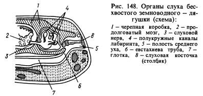
Of the sense organs, the organ of hearing has undergone the most significant changes. The transmission of sound waves from an aquatic environment to animal tissues, which are also saturated with water and have approximately the same acoustic properties as water, occurs much better than from air. Sound waves propagating in the air are almost entirely reflected from the surface of the animal and only about 1% of the energy of these waves penetrates its body. In this regard, amphibians developed, in addition to the labyrinth, or inner ear, a new section of the hearing organ - the middle ear. It is (Fig. 148) a small cavity filled with air, communicating with the oral cavity through the Eustachian tube and closed from the outside by a thin, elastic eardrum. In the middle ear there is an auditory plate (or column), which at one end rests against the eardrum, and at the other against a window covered with a film and leading into the cranial cavity, where there is a labyrinth surrounded by perilymph. The pressure inside the middle ear is equal to atmospheric pressure and the eardrum can vibrate under the influence of sound air waves, the impact of which is further transmitted through the auditory ossicle and perilymph to the walls of the labyrinth and is perceived by the endings of the auditory nerve. The cavity of the middle ear was formed from the first gill slit, and the column was formed from the hyomandibular bone (hyomandibular bone) located near the slit, which suspended the visceral part of the skull to the brain where the labyrinth was located behind the ear bones.
The eyes are covered with movable eyelids, which protect the organs of vision from drying out and clogging. Thanks to changes in the shape of the cornea and lens, amphibians see further than fish. Amphibians perceive small temperature changes well. They are sensitive to the effects of various substances dissolved in water. Their olfactory organ reacts to irritations caused by gaseous substances. Thus, the sensory organs of amphibians have undergone a number of changes in connection with the transition to living on land. Larvae and adult animals that live constantly in water have, like fish, lateral line organs.
Amphibians are characterized by rather complex instinctive actions, especially during the breeding season. For example, the male midwife toad, which lives in Russia in western Ukraine, wraps “cords” of eggs around its hind limbs and hides in secluded places on the shore until the tadpoles develop. After 17-18 days, the male returns to the water, where the tadpoles hatch. This is a kind of instinct to protect offspring. Even more complex instincts are known in a number of tropical tailless amphibians. Amphibians also have conditioned reflexes, but they are developed with great difficulty.
Motor system and skeleton. The muscular system, in connection with various adaptations to life on land (the development of land-type limbs, the emergence of a movable joint between the head and the body, etc.) underwent radical transformations, although it retained many of the features inherent in fish. The muscular system of fish is very uniform and mainly consists of similar lateral muscle segments. In amphibians, the muscular system has become more differentiated, consisting of a variety of muscles (Fig. 149). Amphibians laid the foundations of the muscular system, which later developed and became more complex in real land vertebrates - reptiles, birds and mammals. This also applies to the skeleton.
The skull of amphibians has many cartilaginous elements, which is probably explained by the need to lighten body weight due to a semi-terrestrial lifestyle. The skull contains many bones listed in the description of the skull of higher fish, including the parasphenoid characteristic only of fish and amphibians (Fig. 150). Since the hyomandibular bone has become an auditory bone, the role of the pendant is played by the quadrate bone. Due to the loss of the gill apparatus in adulthood, the gill arches are reduced and only their modified remains are preserved. The hyoid arch changes greatly and is partially reduced. The skull of amphibians is very wide, which is partly due to the characteristics of their breathing. The lower jaw, like that of bony fishes, consists of several bones.
The vertebral column (Fig. 150) in tailless animals is very short and ends in a long bone - the urostyle, formed from the rudiments of the caudal vertebrae. In tailed amphibians, the caudal section of the vertebral column consists of a number of vertebrae. In these amphibians, the tail plays a significant role in movement: in water it is used for swimming, on land it is used to maintain balance. The ribs are poorly developed (in caudate amphibians) or reduced, and their remains are fused with the transverse processes of the vertebrae (in other amphibians); ancient amphibians had ribs. Their reduction in modern forms is explained by the need to lighten the body weight (which greatly increased during the transition from the aquatic environment to the air) in these vertebrates, which are not yet sufficiently adapted to movement on land. Due to the reduction of the ribs, amphibians do not have a chest. The first vertebra is structured differently than in fish: it has two articular sockets for articulation with the two occipital condyles of the skull, due to which the head of amphibians has become mobile.
The skeleton of the forelimb (Fig. 150) consists of the humerus, two forearm bones - the radius and the ulna, carpal bones, metacarpal bones and phalanges of the fingers. The skeleton of the hind limb (Fig. 150) consists of the thigh, two bones of the lower leg - the tibia and fibula, tarsal bones, metatarsal bones and phalanges of the fingers. Consequently, the similarity in the structure of both pairs of limbs, despite some differences in their functions, is very great. Initially, the front and hind legs were five-toed; modern amphibians may have fewer toes. The hind limbs of many tailless amphibians are also used for swimming, and therefore they are elongated, and the fingers are connected by swimming membranes.
The limb girdles are much better developed than those of fish. The shoulder girdle consists of bone and cartilaginous elements: scapula, clavicle, crow bone (coracoid), etc. (Fig. 150). The clavicles and coracoids are connected to the sternum, which also includes bone and cartilaginous elements. The head of the humerus articulates with the shoulder girdle. The posterior girdle of the limbs, or pelvis, consists of three bones: the ilium, the pubis and the ischium (Fig. 150). The large acetabulum formed by these bones serves for articulation with the head of the femur. The pelvis is connected to one vertebra - the sacral one, thanks to which the hind legs, unlike the ventral fins of fish, received quite strong support.
Circulatory system. In amphibian larvae that live in water and breathe with gills, the circulatory system is basically similar to the circulatory system of fish, but in adult animals leading a terrestrial lifestyle, it changes significantly due to the replacement of gill respiration with pulmonary respiration, increased skin respiration, and the development of the limbs of land animals. type and other body changes. The heart (see Fig. 134, B, 151) consists of three chambers: the right and left atria and one ventricle. Departs from the right side of the latter conus arteriosus(it was also present in fish, the ancestors of amphibians), from which four pairs of arteries originate: the first pair - carotid arteries, carrying blood to the head, the second and third pairs are vessels connecting to form the largest vessel of the body - aorta, the branches of which are directed to different parts of the body, the fourth pair - pulmonary arteries, which are then divided into independent cutaneous and pulmonary arteries.
From the lungs, oxygenated blood flows through the pulmonary veins into the left atrium, and blood, saturated in all parts of the body with carbon dioxide, flows into the anterior vena cava in the anterior part of the body, and into the posterior vena cava in the posterior part of the body (Fig. 152 ). Both vena cavae empty into venous sinus, from where blood (saturated with carbon dioxide) flows into the right atrium. From both atria, blood enters the single ventricle of the heart. The inner surface of the ventricle has depressions and therefore the blood in it does not have time to completely mix: in the left part there is blood saturated with oxygen, in the right part there is blood saturated with carbon dioxide, and in the middle part it is mixed. Since the arterial cone begins on the right side of the ventricle, the first portion of blood entering it (i.e., the arterial cone) will be venous, it is sent to the most posterior arteries - the pulmonary ones.
The mixed blood that then flows into the arteries that form the aorta, and through the branches of the latter into all parts of the body. Oxygenated blood from the left side of the ventricle is sent to the carotid arteries. To this it must be added that blood, saturated with oxygen in the skin, enters, as noted above, through the anterior vena cava and venous sinus into the right atrium and thus dilutes the venous blood located there, which is then pushed into the vessels that form the aorta. Consequently, thanks to the devices described above, as well as others not described here, different parts of the body receive blood unequally saturated with oxygen. In Fig. 152 shows the main arterial and venous vessels of amphibians.
In amphibians, due to the strong development of the limbs and greater body dissection than in fish, the network of blood vessels has changed significantly. Many new vessels appeared that were absent in fish, and a system of vessels characteristic of terrestrial vertebrates emerged. At the same time, it should be remembered that the circulatory system of amphibians is much simpler than that of higher vertebrates.
Respiratory system. Almost all amphibians have lungs (see Fig. 151; 153). These organs still have a very simple structure and are thin-walled sacs, in the walls of which a rather dense network of blood vessels branches. Since the inner wall of the lungs is almost smooth, their surface area is relatively small. The trachea is almost undeveloped and the lungs are connected directly to the larynx. Since amphibians do not have a chest (see above), the act of breathing is ensured by the work of the muscles of the oral cavity. Inhalation occurs as follows. With open nostrils (which, unlike the nostrils of fish, are through, i.e., in addition to the external nostrils there are also internal nostrils - choanae) and the mouth is closed, the bottom of the large oral cavity is pulled back and air enters it. Then the nostrils are closed with special valves, the bottom of the mouth is raised and air is forced into the lungs. Exhalation occurs as a result of contraction of the abdominal muscles.
Amphibians receive a significant amount of oxygen through the skin and mucous membranes of the oral cavity. Some species of salamanders have no lungs at all and all gas exchange occurs through the skin. However, the skin can only perform breathing functions if it is moist. Therefore, it is impossible for amphibians to live in conditions where air humidity is insufficient for them. Larvae living in water breathe through gills (first external, then internal) and skin. Some tailed amphibians that constantly live in water have gills that remain throughout their lives. Thus, in terms of breathing methods, amphibians are still close to fish.
Excretory system. The kidneys (see Fig. 136, A, B; Fig. 151), like those of fish, are trunk. Wolffian canals empty into the cloaca. The bladder opens there, where urine accumulates. Removal of dissimilation products also occurs through the skin and lungs.
Digestive system. The oral cavity is very wide. A number of species (mainly tailed amphibians) have many small, uniform, primitively arranged teeth that sit on the jaws, vomer, palatine and other bones and serve only to hold prey. In most species (mainly tailless amphibians), teeth are partially or completely reduced, but their tongue develops greatly. The latter in frogs is attached at the front end and can be thrown far forward with the rear end to catch prey. It is very sticky and well adapted to perform this function. In species that constantly live in water, the tongue is usually reduced. The capture of prey in such amphibians is carried out by the jaws.
The digestive tube (see Fig. 151) is relatively short and consists of the pharynx, esophagus, stomach, small intestine and a very small rectum (colon). The posterior part of the rectum is the cloaca; through it, in addition to feces, urine and sexual products are excreted. Salivary glands, which are absent in fish, flow into the oral cavity. The secretion of these glands serves mainly to moisten food. The salivary glands are very poorly developed in species that live in water, and much better in terrestrial ones. The liver is large; The pancreas is well defined. The food of adult amphibians is mainly animal (insects, small vertebrates, etc.). Tadpoles of tailless amphibians are mostly herbivorous.

Reproduction. The male gonads (testes) lie near the kidneys (see Fig. 151, B). Their ducts open into the tubules of the anterior part of the kidneys (see Fig. 136, A) and the seed is excreted, like urine, through the Wolffian canals. The female gonads (ovaries) grow greatly during the spawning period. The eggs exit through very long Müllerian canals (see Fig. 136, B). The latter do not have a direct connection with the ovaries and the ripening eggs enter through the body cavity into the funnels of the Müllerian canals.
Fertilization in most cases occurs in water. In many amphibians, this is preceded by the rapprochement of the male and female: the male clasps the female from behind, presses his forelimbs on her abdominal wall and thereby facilitates the release of eggs into the water, which he immediately fertilizes. Thus, in the presence of sexual intercourse, fertilization occurs outside the female’s body. In a minority of species (for example, newts), the male releases the seed in a special sac (spermatophore), which the female immediately captures with the edges of the cloaca. In this case, there is no sexual intercourse, but fertilization is internal. Finally, in some species the male inserts seed into the female's cloaca using his protruding cloaca.
In many species, sexual dimorphism is well expressed (in color, the structure of the front legs with which males hold females, and other characteristics). Males of a number of species can produce very loud sounds due to the amplification of these sounds by vocal sacs - resonators.
Development. Amphibian development usually occurs in water. From fertilized eggs, larvae (tadpoles) develop, which have a fish-like shape. They breathe through gills and their internal structure resembles fish. During the growth period, transformation (metamorphosis) of tadpoles occurs: first, their hind legs grow, then their front legs, gills and tail atrophy (in tailless animals), lungs develop, major changes occur in the circulatory system, etc.
Origin. Amphibians, as explained above (p. 296), descended from lobe-finned fish. The paired fins of ancient lobe-finned fish, from which the articulated limbs of terrestrial vertebrates developed, were short and wide, they included many small bone elements, not connected by joints, located in several (at least eight) transverse rows. The girdles on which the fins rested were relatively poorly developed (especially the pelvic girdle). Due to the transformation of fins into terrestrial limbs, significant changes occurred in the skeleton.
Firstly, many bone elements were reduced: in the first proximal rows there was only one bone left, in the front leg - the shoulder, in the back - the femur; in the second rows - two bones each, in the front leg - the radius and ulna, in the back - the tibia and fibula; in the next two rows, five bones remained, forming the carpus in the front leg and the tarsus in the back leg; in the next row, the remaining five bones were included in the metacarpus in the front leg, and the metatarsals in the rear leg; the remaining three rows with five bones each became the phalanges of the fingers. Reducing the number of bones contributed to increased strength of the legs.
Secondly, the bones of the first two rows (i.e., shoulder and forearm, thigh and lower leg) were greatly lengthened, which was very important for enhancing the speed of movement.
Thirdly, joints developed between the listed bones, that is, the limbs became articulated, which is the most important condition for their work.
Fourthly, the limb belts have been strengthened (see above for the description of the belts). In parallel with these changes, profound changes occurred in the nervous, muscular and vascular systems of the legs. The changes in other organ systems that occurred during the transformation of lobe-finned fish into amphibians are described in the general characteristics of the latter.
The most ancient amphibians were stegocephalians (Fig. 154), which were numerous in the Carboniferous period and finally became extinct in the Triassic. They lived along the banks of reservoirs and spent a lot of time in the water. The head of these animals was covered with scutes, which explains their name (stegocephals - covered-headed). Their skeleton had many primitive features: the bone elements of the legs were small and slightly different in size, the vertebrae were biconcave, the girdles of the limbs were poorly developed, etc. Modern groups of amphibians originated from them.
Brain frogs, like other amphibians, are characterized by the following features compared to fish:
a) progressive development of the brain, expressed in the separation of the paired hemispheres by a longitudinal fissure and the development of the gray matter of the ancient cortex (archipallium) in the roof of the brain;
b) weak development of the cerebellum;
c) weak expression of the bends of the brain, due to which the intermediate and middle sections are clearly visible from above.
Diamond brain(rhombencephalon)
Medulla oblongata (myelencephalon, medulla oblongata), into which the spinal cord passes cranially, it differs from the latter in its greater width and the departure from its lateral surfaces of the large roots of the posterior cranial nerves. On the dorsal surface of the medulla oblongata there is diamond-shaped fossa (fossa rhomboidea), accommodating fourth cerebral ventricle (ventriculus quartus). On top it is covered with a thin vascular cap, which is removed along with the meninges. The ventral fissure, a continuation of the ventral fissure of the spinal cord, runs along the ventral surface of the medulla oblongata. The medulla oblongata contains two pairs of cords (bundles of fibers): the lower pair, separated by the ventral fissure, are motor, the upper pair are sensory. The medulla oblongata contains the centers of the jaw and sublingual apparatus, the organ of hearing, as well as the digestive and respiratory systems.
Cerebellum located in front of the rhomboid fossa in the form of a high transverse ridge as an outgrowth of its anterior wall. The small size of the cerebellum is determined by the small and uniform mobility of amphibians - in fact, it consists of two small parts, closely connected with the acoustic centers of the medulla oblongata (these parts are preserved in mammals as fragments of the cerebellum (flocculi)). The body of the cerebellum - the center of coordination with other parts of the brain - is very poorly developed.
Midbrain(mesencephalon) when viewed from the dorsal side, it is represented by two typical optic lobes (lobus opticus s. tectum opticus), having the appearance of paired ovoid elevations forming the upper and lateral parts of the midbrain. The roof of the optic lobes is formed by gray matter - several layers of nerve cells. The tectum in amphibians is the most significant part of the brain. The optic lobes contain cavities that are lateral branches cerebral (Sylvii) aqueduct (aquaeductus cerebri (Sylvii), connecting the fourth cerebral ventricle with the third.
The bottom of the midbrain is formed by thick bundles of nerve fibers - cerebral peduncles (cruri cerebri), connecting the forebrain with the medulla oblongata and spinal cord.
Forebrain(prosencephalon) consists of the diencephalon and telencephalon, lying sequentially.
Diencephalon visible from above as a rhombus, with sharp angles directed to the sides.
Parts of the diencephalon lie around a vertically located wide fissure third cerebral ventricle (ventriculus tertius). Lateral thickening of the walls of the ventricle - visual cusps or thalamus. In fish and amphibians, the thalamus is of secondary importance (as coordinating sensory and motor centers). The membranous roof of the third cerebral ventricle - the epithalamus or epithalamus - does not contain neurons. It contains the superior medullary gland - pineal gland (epiphisis). In amphibians, the pineal gland already serves as a gland, but has not yet lost the features of the parietal organ of vision. In front of the epiphysis, the diencephalon is covered with a membranous roof, which orally turns inward and passes into the anterior choroid plexus (choroid tectum of the third ventricle), and then into the endplate of the diencephalon. Inferiorly the ventricle narrows, forming pituitary funnel (infundibulum), the inferior medullary gland is attached to it caudoventrally - pituitary gland (hypophisis). In front, on the border between the bottom of the terminal and intermediate sections of the brain, there is chiasma nervorum opticorum). In amphibians, most of the fibers of the optic nerves are not retained in the diencephalon, but go further to the roof of the midbrain.
Telencephalon its length is almost equal to the length of all other parts of the brain. It consists of two parts: the olfactory brain and two hemispheres, separated from each other sagittal (arrow-shaped) fissure (fissura sagittalis).
Hemispheres of the telencephalon (haemispherium cerebri) occupy the posterior two-thirds of the telencephalon and hang over the anterior part of the diencephalon, partially covering it. There are cavities inside the hemispheres - lateral cerebral ventricles (ventriculi lateralis), caudally communicating with the third ventricle. In the gray matter of the cerebral hemispheres of amphibians, three areas can be distinguished: dorsomedially there is the old cortex or hippocampus (archipallium, s. hippocampus), laterally - ancient bark(paleopallium) and ventrolaterally - the basal ganglia, corresponding striata (corpora striata) mammals. The striatum and, to a lesser extent, the hippocampus are correlative centers, the latter associated with olfactory function. The ancient cortex is an exclusively olfactory analyzer. On the ventral surface of the hemispheres, grooves are noticeable, separating the striatum from the ancient cortex.
Olfactory brain (rhinencephalon) occupies the anterior part of the telencephalon and forms olfactory lobes (bulbs) (lobus olfactorius), soldered in the middle with each other. They are separated from the hemispheres laterally by the marginal fossa. The olfactory lobes anteriorly contain the olfactory nerves.
10 pairs extend from the frog's brain cranial nerves. Their formation, branching and zone of innervation are not fundamentally different from those in mammals
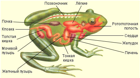
137. Look at the pictures. Write the names of the frog's body parts. What organs are located on her head? Write their names.
138. Study the table "Class Amphibians. Structure of a frog." Look at the drawing. Write the names of the internal organs of the frog, indicated by numbers.
3. stomach
4. pancreas
5. testis
7. Bladder
8. colon
9. small intestine
10. gallbladder
11. liver
139. Describe the structure of the amphibian brain.
The brain of amphibians has more progressive features, larger forebrain sizes, and complete separation of the hemispheres. The midbrain is relatively small. The cerebellum is small because amphibians have monotonous movements. There are 10 cranial nerves leaving the brain.
Divisions of the brain: anterior, middle, cerebellum, medulla oblongata, intermediate.
140. Study the table "Class Amphibians. Structure of a frog." Look at the drawing. Label the parts of the frog's skeleton indicated by numbers.

2. shoulder blade
4. forearm
9. urostyle
10. spine
141. Look at the drawing. Write the names of the frog's digestive system, indicated by numbers. How is digestion carried out in a frog?

1. mouth opening
2. esophagus
3. stomach
4. intestines
All amphibians feed only on mobile food. At the bottom of the oral cavity is the tongue. When catching prey, it is thrown out of the mouth and the prey is attached to it. The upper jaw has teeth that serve only to hold prey. When swallowing, the eyeballs help push food into the esophagus.
The ducts of the salivary glands open into the oropharynx. From the oropharynx, food enters the stomach through the esophagus, and from there into the duodenum. The ducts of the liver and pancreas open here. Digestion occurs in the stomach and duodenum. The small intestine passes into the rectum, which forms an extension - the cloaca.
142. Draw a diagram of the structure of a frog’s heart. Which blood is called arterial and which is called venous?
Arterial blood comes from the lungs and is rich in oxygen. Venous - to the lungs.

143. Describe the process of reproduction and development of a frog. Indicate the similarities and differences in the reproduction of amphibians and fish.
Amphibians breed in small, well-warmed areas of water bodies. The reproductive organs of males are the testes, and the reproductive organs of females are the ovaries. Fertilization is external.
Development of the frog: egg - tadpole at the moment of hatching - development of fin folds and external gills - stage of maximum development of external gills - stage of disappearance of external gills - stage of appearance of the hind limbs - stage of dismemberment and mobility of the hind limbs - stage of release of the forelimbs, metamorphosis of the oral apparatus and beginnings tail resorption - stage of reaching land.
144. Fill out the table.
The structure and significance of the frog's sense organs.
Frog sense organs Structural features Meaning Eyes There are upper (leathery) and lower (transparent) movable eyelids, a nictitating membrane. There is a gland, the secretion of which moistens the cornea and protects it from drying out. The cornea is convex. The lens has the shape of a biconvex lens. Many people have developed color vision Vision Hearing organ Inner ear, middle ear. Externally, the auditory opening is closed by the eardrum, connected to the auditory ossicle - the stapes. Hearing Organ of balance Inner ear Orientation Olfactory organ Paired olfactory sacs. Their walls are lined with olfactory epithelium. They open outward with the nostrils, and into the oropharynx with the chaons. Odor perception Organ of touch Leather Perception of irritation Lateral line organ Lateral line in larvae Allows you to feel the flow of water
Body Amphibians: Divided into head, trunk and five-fingered limbs. Tailed amphibians have a tail.
Reptiles: Divided into head, neck, trunk, tail and five-fingered limbs.
Skin of Amphibians: Thin, devoid of scales, but has a large number of glands that secrete mucus.
Reptiles: Dry, devoid of glands and covered with horny scales that protect the body from drying out. Scales restrain growth, so molting is typical for reptiles.
Spine
Amphibians: 4 sections: cervical, trunk, sacral and caudal. The ribs are reduced and are absent in anurans. The muscles do not have a segmental structure and are represented by differentiated muscle groups.
Reptiles: 5 sections: cervical, thoracic, lumbar, sacral and caudal. There are ribs, a sternum and a rib cage. The parts of the skeleton of the limbs are the same as those of amphibians. The muscles are more differentiated.
Digestive system of Amphibians: The digestive tube is divided into anterior, middle and posterior sections. The stomach is isolated. The expansion of the colon forms a cloaca. Digestive glands are developed.
Reptiles: Oral cavity, pharynx, esophagus, stomach, small and large intestines. At the border of the large and small intestines there is the rudiment of the cecum. The large intestine opens into the cloaca. Digestive glands are developed.
Excretory organs Amphibians: Paired trunk ureters and bladder, which opens into the cloaca.
Reptiles: Secondary (pelvic) kidneys, ureters, bladder (opens into the cloaca).
Circulatory system
Amphibians: The heart is three-chambered. Two circles of blood circulation. Mixed blood flows through the vessels of the systemic circle, and the brain is supplied with arterial blood. Amphibians are poikilothermic animals.
Reptiles: The heart is three-chambered, but the ventricle has an incomplete septum. Two circles of blood circulation.
Respiratory organs: Adult amphibians have lungs; larvae have gills. Additionally, the skin is involved in breathing.
Reptiles: Lungs. They are stretchable bags with an internal mesh which has a network of crossbars that increase the surface. The posterior end of the trachea branches into two bronchi, which enter the lungs.
Answer