What vessels flow into the right atrium of the heart. Chambers of the heart
The heart has a complex structure and performs no less complex and important work. Contracting rhythmically, it ensures blood flow through the vessels.
The heart is located behind the sternum, in the middle section chest cavity and is almost completely surrounded by lungs. It may move slightly to the side as it hangs freely on the blood vessels. The heart is located asymmetrically. Its long axis is inclined and forms an angle of 40° with the axis of the body. It is directed from top to right, forward, down to the left, and the heart is rotated so that its right section is tilted more forward, and the left one – back. Two-thirds of the heart is to the left of the midline and one-third (the vena cava and right atrium) is to the right. Its base is turned towards the spine, and its apex is facing the left ribs, to be more precise, the fifth intercostal space.

Sternocostal surface the hearts are more convex. It is located behind the sternum and cartilages of the III-VI ribs and is directed forward, upward, and to the left. The transverse coronary groove runs along it, which separates the ventricles from the atria and thereby divides the heart into top part, formed by the atria, and the lower one, consisting of the ventricles. Another groove of the sternocostal surface - the anterior longitudinal - runs along the border between the right and left ventricles, with the right one forming the largest part of the anterior surface, the left one the smaller one.
Diaphragmatic surface flatter and adjacent to the tendon center of the diaphragm. Along this surface runs a longitudinal posterior groove, separating the surface of the left ventricle from the surface of the right. In this case, the left one makes up the majority of the surface, and the right one makes up the smaller part.
Anterior and posterior longitudinal grooves they merge at their lower ends and form a cardiac notch to the right of the cardiac apex.
There are also side surfaces located on the right and left and facing the lungs, which is why they are called pulmonary.
Right and left edges hearts are not the same. The right edge is more pointed, the left is more blunt and rounded due to the thicker wall of the left ventricle.
The boundaries between the four chambers of the heart are not always clearly defined. The landmarks are the grooves in which the blood vessels of the heart are located, covered with fatty tissue and the outer layer of the heart - the epicardium. The direction of these grooves depends on how the heart is located (obliquely, vertically, transversely), which is determined by the body type and the height of the diaphragm. In mesomorphs (normosthenics), whose proportions are close to the average, it is located obliquely, in dolichomorphs (asthenics) with a thin physique - vertically, in brachymorphs (hypersthenics) with wide short forms - transversely.

The heart seems to be suspended by the base on large vessels, while the base remains motionless, and the apex is in a free state and can move.
Structure of heart tissue
The heart wall is made up of three layers:
- Endocard – inner layer epithelial tissue lining the cavities of the heart chambers from the inside, accurately repeating their relief.
- The myocardium is a thick layer formed by muscle tissue (striated). The cardiac myocytes of which it consists are connected by many bridges that link them into muscle complexes. This muscle layer ensures the rhythmic contraction of the chambers of the heart. The myocardium is thinnest at the atria, the greatest is at the left ventricle (about 3 times thicker than the right), since it needs more force to push blood into big circle blood circulation, in which the resistance to flow is several times greater than in the small one. The atrial myocardium consists of two layers, the ventricular myocardium - of three. The atrial myocardium and ventricular myocardium are separated by fibrous rings. The conduction system that provides rhythmic contraction of the myocardium is one for the ventricles and atria.
- The epicardium is the outer layer, which is the visceral petal of the heart sac (pericardium), which is a serous membrane. It covers not only the heart, but also primary departments pulmonary trunk and aorta, as well as the final sections of the pulmonary and vena cava.

Anatomy of the atria and ventricles
The cardiac cavity is divided by a septum into two parts - right and left, which do not communicate with each other. Each of these parts consists of two chambers - the ventricle and the atrium. The septum between the atria is called the interatrial septum, and the septum between the ventricles is called the interventricular septum. Thus, the heart consists of four chambers - two atria and two ventricles.
Right atrium
In shape it looks like an irregular cube, in front there is additional cavity, called the right ear. The atrium has a volume of 100 to 180 cubic meters. cm. It has five walls, 2 to 3 mm thick: anterior, posterior, superior, lateral, medial.
The superior vena cava (from above, behind) and the inferior vena cava (from below) flow into the right atrium. On the lower right is the coronary sinus, where the blood of all the cardiac veins drains. Between the openings of the superior and inferior vena cava there is an intervenous tubercle. In the place where the inferior vena cava flows into the right atrium, there is a fold of the inner layer of the heart - the valve of this vein. The sinus of the vena cava is the posterior dilated section of the right atrium, into which both of these veins flow.
The chamber of the right atrium has a smooth internal surface, and only in the right appendage with the adjacent anterior wall the surface is uneven.
Many pinpoint openings of the small veins of the heart open into the right atrium.
Right ventricle
It consists of a cavity and an arterial cone, which is a funnel directed upward. The right ventricle has the shape of a triangular pyramid, the base of which faces upward and the apex faces downward. The right ventricle has three walls: anterior, posterior, medial.
The front is convex, the back is flatter. The medial is the interventricular septum, consisting of two parts. The larger one, the muscular one, is located at the bottom, the smaller one, the membranous one, is at the top. The pyramid faces the atrium with its base and has two openings: posterior and anterior. The first is between the cavity of the right atrium and the ventricle. The second goes into the pulmonary trunk.
Left atrium
It has the appearance of an irregular cube, is located behind and adjacent to the esophagus and the descending aorta. Its volume is 100-130 cubic meters. cm, wall thickness – from 2 to 3 mm. Like the right atrium, it has five walls: anterior, posterior, superior, literal, medial. The left atrium continues anteriorly into an additional cavity called the left appendage, which is directed towards the pulmonary trunk. Four enter the atrium pulmonary veins(back and top), in the holes of which there are no valves. The medial wall is the interatrial septum. The inner surface of the atrium is smooth, the pectineus muscles are present only in the left appendage, which is longer and narrower than the right one, and is noticeably separated from the ventricle by an interception. It communicates with the left ventricle via the atrioventricular orifice.
Left ventricle
It is shaped like a cone, the base of which faces upward. The walls of this chamber of the heart (anterior, posterior, medial) have the greatest thickness - from 10 to 15 mm. There is no clear boundary between the front and back. At the base of the cone are the openings of the aorta and the left atrioventricular opening.
The round opening of the aorta is located in front. Its valve consists of three valves.
Heart size
The size and weight of the heart differs among different people. The average values are as follows:
- length is from 12 to 13 cm;
- greatest width – from 9 to 10.5 cm;
- anteroposterior size – from 6 to 7 cm;
- weight in men - about 300 g;
- weight in women is about 220 g.

Functions of the cardiovascular system and heart
The heart and blood vessels make up the cardiovascular system, the main function of which is transport. It consists of supplying tissues and organs with nutrition and oxygen and returning metabolic products.
The heart acts as a pump - it ensures continuous circulation of blood in the circulatory system and delivery of nutrients and oxygen to organs and tissues. When stressed or physical activity his work is immediately restructured: the number of layoffs increases.
The work of the heart muscle can be described as follows: its right part (venous heart) receives waste blood saturated with carbon dioxide from the veins and gives it to the lungs to be saturated with oxygen. From the lungs, O2-enriched blood is directed to the left side of the heart (arterial) and from there is forcefully pushed into the bloodstream.
The heart produces two circles of blood circulation - large and small.
The large one supplies blood to all organs and tissues, including the lungs. It begins in the left ventricle and ends in the right atrium.
The pulmonary circulation produces gas exchange in the alveoli of the lungs. It begins in the right ventricle and ends in the left atrium.
Blood flow is regulated by valves: they prevent it from flowing into reverse direction.
The heart has such properties as excitability, conductivity, contractility and automaticity (excitation without external stimuli under the influence internal impulses).
Thanks to the conduction system, sequential contraction of the ventricles and atria occurs, and the synchronous inclusion of myocardial cells in the contraction process.
Rhythmic contractions of the heart ensure a portioned flow of blood into the circulatory system, but its movement in the vessels occurs without interruption, which is due to the elasticity of the walls and the resistance to blood flow that occurs in small vessels.
The circulatory system has a complex structure and consists of a network of vessels for different purposes: transport, shunting, exchange, distribution, capacitance. There are veins, arteries, venules, arterioles, capillaries. Together with the lymphatics, they maintain the constancy internal environment in the body (pressure, body temperature, etc.).
Arteries move blood from the heart to the tissues. As they move away from the center, they become thinner, forming arterioles and capillaries. Arterial bed circulatory system transports necessary substances to the organs and maintains constant pressure in the vessels.

The venous bed is more extensive than the arterial bed. Veins move blood from tissues to the heart. Veins are formed from venous capillaries, which, merging, first become venules, then veins. They form large trunks near the heart. Distinguish superficial veins, located under the skin, and deep, located in the tissues near the arteries. The main function of the venous part of the circulatory system is the outflow of blood, rich in products metabolism and carbon dioxide.
For rate functionality of cardio-vascular system and the permissibility of loads, special tests are carried out, which make it possible to assess the performance of the body and its compensatory capabilities. Functional tests cardiovascular system are included in the medical physical examination to determine the degree of fitness and general physical fitness. The assessment is based on such indicators of the functioning of the heart and blood vessels as blood pressure, pulse pressure, blood flow speed, minute and stroke volumes of blood. Such tests include Letunov's tests, step tests, Martinet's test, Kotov's - Demin's test.
The heart begins to beat from the fourth week after conception and does not stop until the end of life. It does a gigantic job: per year it pumps about three million liters of blood and makes about 35 million heartbeats. At rest, the heart uses only 15% of its resource, and under load – up to 35%. Behind average duration During its lifetime it pumps about 6 million liters of blood. Another interesting fact: The heart supplies blood to 75 trillion cells in the human body, excluding the cornea of the eyes.
called blood circulation. Through circulation, blood communicates
all organs of the human body, there is a supply of nutrients and
oxygen, removal of metabolic products, humoral regulation and etc.
Blood moves through blood vessels. They represent
elastic tubes of different diameters. The main circulatory organ is
heart - hollow muscular organ making rhythmic contractions.
Thanks to its contractions, blood moves in the body. Doctrine of
regulation of blood circulation developed by I.P. Pavlov.
There are 3 types of blood vessels: arteries, capillaries and veins.
Arteries- vessels through which blood flows from the heart to the organs. They have
thick walls consisting from 3 layers:
Outer layer ( adventitia) – connective tissue;
- average ( media) – consists of smooth muscle tissue and contains
connective tissue elastic fibers. Cutting this shell
accompanied by a decrease in the lumen of blood vessels;
Internal ( intimate) – formed by connective tissue and from the side
The lumen of the vessel is lined with a layer of flat endothelial cells.
The arteries are located deep under the muscle layer and are reliably protected from
damage. As they move away from the heart, the arteries branch into smaller vessels,
and then to the capillaries.
Depending on the blood supply to the organs and tissues, arteries are divided into:
1. Parietal ( wall) - blood supply to the walls of the body.
2. Visceral ( visceral) – blood supply to internal organs.
Before an artery enters an organ, it is called an organ; upon entering an organ, it is called an organ.
intraorgan. Depending on the development of different layers of the artery wall
are divided into vessels:
- muscular type – they have a well-developed middle shell and fibers
arranged spirally like a spring;
Mixed ( muscular-elastic) type – approximately equal in the walls
number of elastic and muscle fibers(carotid, subclavian);
- elastic type in which the outer shell is thinner than the inner.
These are the aorta and pulmonary trunk, into which blood flows under high pressure.
In children, the diameter of the arteries is larger than in adults. In newborns, arteries
predominantly elastic type, arteries of the muscular type are not yet developed.
Capillaries are the smallest blood vessels with
clearance from 2 to 20 microns. The length of each capillary does not exceed 0.3 mm. Their
the quantity is very large, so for 1mm2 of fabric there are several hundred
capillaries. The total lumen of the capillaries of the whole body is 500 times larger than the lumen of the aorta.
In a resting state of the organ most of capillaries do not function and current
the blood in them stops. The capillary wall consists of one layer
endothelial cells. The surface of the cells facing the lumen of the capillary
uneven, wrinkles form on it. Metabolism between blood and tissues
occurs only in capillaries. Arterial blood along the capillaries
turns into venous, which collects first in postcapillaries, and then in
Distinguish capillaries:
1. Feeding– provide the organ with nutrients and O2, and
2. Specific– create the opportunity for the body to perform its function
(gas exchange in the lungs, excretion in the kidneys).
Vienna- These are vessels through which blood flows from organs to the heart. They,
like arteries, they have three-layer walls, but contain less elastic and
muscle fibers, therefore less elastic and easily collapse. Veins have
valves that open with blood flow. This promotes blood movement in
one direction. The movement of blood in one direction in the veins is facilitated by
not only the semilunar valves, but also the pressure difference in the vessels and contractions
muscle layer of veins.
Each area or organ receives its blood supply from several vessels.
There are:
1. Main vessel- the biggest.
2. Additional ( collateral) is a lateral vessel that carries out
roundabout blood flow.
3. Anastomosis- This is the third vessel that connects the other 2. Otherwise
called connecting vessels.
There are also anastomoses between the veins. Termination of current in one vessel
leads to increased blood flow through collateral vessels and anastomoses.
CIRCULATION PATTERN
Blood circulation is necessary to nourish the tissues where metabolism takes place
substances through the walls of capillaries. Capillaries constitute the main part
microvasculature, in which microcirculation of blood occurs and
Microcirculation- this is the movement of blood and lymph in the microscopic
parts of the vascular bed. The microcirculatory bed according to V.V. Kupriyanov includes
5 links:
1. Arterioles- the smallest parts of the arterial system.
2. Precapillaries– intermediate link between arterioles and true
capillaries.
3. Capillaries.
4. Postcapillaries.
5. Venules.
All blood vessels in the human body make up 2 circles of blood circulation:
small and large.
Lecture 9. LYMPHATIC SYSTEM
It is represented by lymph nodes and lymphatic vessels, in
which lymph circulates.
Lymph in its composition resembles blood plasma, in which suspended
lymphocytes. In the body there is a constant formation of lymph and its outflow through
lymph vessels into veins. The process of lymph formation is associated with metabolism between
blood and tissue.
As blood flows through the blood capillaries, part of its plasma
tissue and makes up tissue fluid. Tissue fluid washes the cells, when
In this case, a constant exchange of substances occurs between the fluid and the cells: in
cells arrive nutrients and oxygen, and vice versa - metabolic products.
Tissue fluid containing metabolic products partially returns to the
blood through the walls of blood vessels. At the same time, another part of the tissue
fluid does not enter the blood, but into the lymph vessels and constitutes lymph. So
way, lymphatic system is an additional outflow system,
complementing the function of the venous system.
Lymph- translucent yellowish liquid formed from
tissue fluid. In its composition it is close to blood plasma, but the proteins in it
less. Lymph contains many leukocytes that enter it from
intercellular spaces and lymph nodes. Lymph flowing from different
organs has a different composition. By lymphatic vessels she enters
circulatory system (about 2 liters per day). Lymph nodes perform protective
drains into the right venous angle. Lymph flows into it from the right half
chest, right upper limb, right half of the head, face and neck.
Can spread through lymphatic vessels along with lymph
pathogenic microbes and particles of malignant tumors.
Along the path of the lymph vessels, lymph nodes are located in some places. By
bringing vessels, lymph flows to the nodes, along related- flows away from them.
Lymph nodes are small round or oblong
Taurus Each node consists of a connective tissue membrane, from which inwards
the crossbars come off. The skeleton of the lymph nodes consists of reticular tissue. Between
the crossbars of the nodules contain follicles in which reproduction occurs
lymphocytes.
Functions lymph nodes:
They are hematopoietic organs
Perform a protective function (pathogenic microbes are retained);
in such cases, the nodes increase in size, become dense and may
palpate.
Lymph nodes are located in groups. Lymph from each organ or area
premature puberty.
THYMUS
Thymus located in the upper part of the anterior mediastinum
directly behind the sternum. It consists of two (right and left) lobes , upper
the ends of which can exit through the upper opening of the chest, and the lower
often extend to the pericardium and occupy the upper interpleural space
triangle. The size of the gland during a person’s life is not the same: its mass varies
a newborn averages 12 g, at 14-15 years old - about 40, at 25 years old - 25 and at 60 years old -
close 15 g . In other words, thymus, having achieved the greatest development
time of puberty, and subsequently gradually decreases.
The thymus gland is of great importance in immune processes, its hormones are up to
the onset of puberty inhibits the function of the gonads, regulates __________ growth
bones (osteosynthesis), etc.
ADRENAL GLAND
Adrenal gland(glandiila suprarenalis) steam room, refers to so
called the adrenal system. Located in the retroperitoneal space -
directly at the upper pole of the kidney. This gland is shaped like a three-
a faceted pyramid, with its apex facing the diaphragm and its base facing the kidney.
Its dimensions in an adult: height 3-6 cm , base diameter about 3 cm
and the width is approximately 4-6 mm , weight – 20 g . On the anterior surface of the gland there are
gate – the place of entry and exit of blood vessels and nerves. Gland covered
connective tissue capsule, which is part of the renal fascia. From-
the sprouts of the capsule penetrate into it through the gate and form, as it were, an organ stroma.
In cross section, the adrenal gland consists of an outer cortex
matter and inner medulla.
The adrenal medulla secretes a group of adrenaline hormones.
of the first series, which stimulate the function of the sympathetic nervous system: narrowing
blood vessels, stimulate the process of breakdown of glycogen in the liver and
etc. Hormones secreted by the adrenal cortex, or
choline-like substances regulate water-salt metabolism and affect the function
gonads.
Lecture 11. STUDY ABOUT THE NERVOUS SYSTEM (NEUROLOGY)
DEVELOPMENT OF THE NERVOUS SYSTEM
Stage 1 - reticular nervous system. At this stage (coelenteric)
the nervous system consists of nerve cells, whose numerous branches
connect to each other in different directions, forming a network. Reflection of this
stage in humans is a network-like structure nervous system digestive
Stage 2 – nodal _________nervous system. At this stage (invertebrate) nervous
cells come together into separate clusters or groups, and from the clusters
cell bodies are obtained ganglia- centers, and from clusters of processes -
nerves. With a segmental structure, nerve impulses arising at any point
bodies, do not spread throughout the body, but spread along transverse trunks in
within this segment. A reflection of this stage is the preservation in humans
primitive features in the structure of the autonomic nervous system.
Stage 3 – tubular nervous system. Such a nervous system (NS) in chordates
(lancelet) arose in the form of a neural tube with segmental segments extending from it
nerves to all segments of the body, including the movement apparatus - the trunk brain. U
In vertebrates and humans, the trunk cord becomes the spinal cord. Phylogeny of NS
determines the embryogenesis of the human NS. The NS is formed in the human embryo at
second or third week intrauterine development. It comes from the outside
germ layer - ectoderm, which forms the medulla. This
the plate deepens, turning into the brain tube. Brain tube
represents the rudiment of the central part of the NS. The rear end of the tube forms
rudiment spinal cord. Front widened end by constriction
divided into 3 primary brain vesicles, from which the brain arises
144
The neural plate initially consists of a single layer of epithelial
cells. During its closure into the brain tube, the number of cells increases
and 3 layers arise:
Internal, from which the epithelial lining of the brain stems
cavities;
The middle one, from which the gray matter of the brain develops (germinal
nerve cells);
External, developing in white matter(nerve cell processes). At
separation of the brain tube from the ectoderm is formed ganglionic plate. From her
spinal nodes develop in the area of the spinal cord, and in the area of the brain
brain - peripheral nerve nodes. Part of the ganglion neural plate goes
on the formation of ganglion nodes) of the autonomous NS, located in the body on
various distances from the central nervous system (CNS).
The walls of the neural tube and the ganglion plate consist of cells:
Neuroblasts from which neurons develop (functional unit
nervous system);
Neuroglial cells are divided into macroglial and microglial cells.
Macroglia cells develop like neurons, but are not capable of conducting
excitation. They perform protective functions, power and contact function
between neurons.
Microglial cells originate from mesenchyme ( connective tissue). Cells
together with blood vessels enter the brain tissue and are phagocytes.
IMPORTANCE OF THE NERVOUS SYSTEM
1. The National Assembly regulates activities various organs, organ systems and everything
body.
2. Connects the whole organism with external environment. All the irritations from
external environment are perceived by the NS using the senses.
3. The NS carries out connections between different organs and systems and
coordinates the activities of all organs and systems, ensuring integrity
body.
4. The human brain is the material basis of thinking and
speech associated with it.
CLASSIFICATION OF THE NERVOUS SYSTEM
The NS is divided into two closely interconnected parts.
Anatomical features
The right atrium is located anterior and to the right relative to the left. Outside it is covered with an epicardium, under which there are thin layer myocardium and the inner layer - endocardium. From the inside of the atrium, the surface is smooth, except for the inner surface of the appendage and part of the anterior wall, where ribbing is noticeable. This ribbing is due to the presence of the pectineus muscles, which are delimited by the border ridge from the rest of the inner surface. The right ear is an additional cavity in the shape of a pyramid.
The appendage functions as a blood reservoir and decompression chamber during ventricular systole. The ear also has a receptor zone, which allows it to take part in the regulation of heart contractions. Not far from the ear, on the anterior wall, there is an atrioventricular opening, through which communication occurs with the ventricle. The medial wall of the atrium plays the role of the interatrial septum. It has an oval fossa, which is closed by a thin connective tissue membrane.

Before birth and during the neonatal period, in its place is the foramen ovale, which takes part in the fetal circulation. After birth, the function of the foramen ovale is lost and it closes, leaving a hole. In a quarter of the population, the opening does not close and an atrial septal defect called the foramen ovale is formed.
In most cases, the defect does not cause any problems, but over time, with large sizes oval window, there is a risk of paradoxical embolism and heart attacks. The foramen ovale also ensures the discharge of blood from the left to the right atrium, which causes the mixing of arterial and venous blood and decreased cardiac output.
2 Emerging vessels

The superior and inferior vena cava are the two largest veins in the body, to which blood flows from all organs and tissues. Along with the vena cava, the smallest veins of the heart and the coronary sinus flow into the right atrium. The smallest veins of the heart open into the atrium along its entire surface. The coronary sinus is a collector of the veins of the heart, which, with the help of an orifice, opens into the atrium cavity between the opening of the inferior vena cava and the atrioventricular opening. The veins draining into the coronary sinus represent the main route for the outflow of venous blood from the heart. After passing through the atrium, it goes to the ventricle.
3 Beginning of the conduction system of the heart
Between the mouth of the superior vena cava and the right ear is the sinoatrial node. It coordinates the work of different parts of the heart, ensuring normal cardiac activity. The sinoatrial node generates impulses and is a first-order pacemaker (70 per minute). From it, the right and left branches of the sinoatrial node go to the myocardium.
4 Physiology and significance in the cardiac cycle

Exactly anatomical features the structures of the atrium ensure continuity and constancy of blood flow even during contraction of the ventricles. A number of factors contribute to constant venous inflow, one of which is thin walls. Thin walls cause stretching of the atrium, as a result of which it does not have time to fill with blood. Due to the thin muscle layer, the right atrium does not completely contract during systole, which ensures transient blood flow from the veins through the atrium into the ventricle.
Since the contractions are quite weak, they do not cause a significant increase in pressure that would impede venous flow or promote the backflow of blood into the veins. Another factor ensuring continuous circulation is the absence of inlet valves at the mouth of the vena cava, the opening of which would require an increase in venous pressure. In addition, the presence of atrial volume receptors plays a significant role in maintaining blood flow.
These are baroreceptors low pressure, which send signals to the hypothalamus when pressure decreases. A decrease in pressure indicates a decrease in blood volume. The hypothalamus responds to this by releasing vasopressin. Summarizing the above, we can conclude that without the right atrium, due to periodic increase pressure during ventricular contraction, blood flow to the heart would be jerky, which would affect overall speed blood circulation in the direction of its reduction.
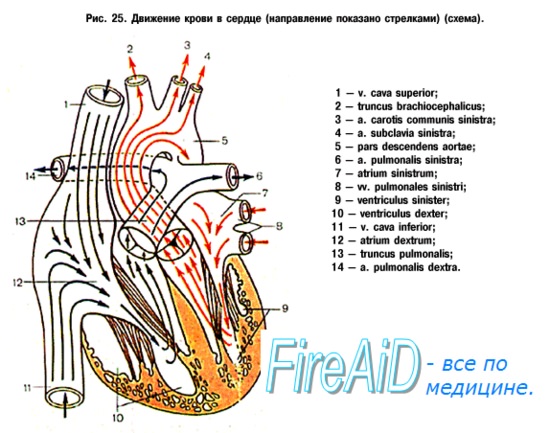
Atria are chambers that receive blood; the ventricles, on the contrary, eject blood from the heart into the arteries. The right and left atria are separated from each other by a septum, as are the right and left ventricles. On the contrary, between the right atrium and the right ventricle there is a communication in the form right atrioventricular orifice, ostium atrioventriculare dextrum; between left atrium and left ventricle - ostium atrioventriculare sinistrum.
Through these openings, blood is directed from the cavities of the atria into the cavities of the ventricles during atrial systole.
Right atrium, atrium dextrum, has the shape of a cube. From behind they pour into it at the top v. cava superior and below v. cava inferior, anteriorly, the atrium continues into the hollow process - the right ear, auricula dextra. The right and left ears cover the base of the aorta and pulmonary trunk. The septum between the atria, septum interatriale, is set obliquely, from the anterior wall it goes back and to the right, so that the right atrium is located on the right and in front, and the left atrium is located on the left and behind. The inner surface of the right atrium is smooth, with the exception of a small area in front and the inner surface of the appendage, where a number of vertical ridges located here are noticeable pectineus muscles, musculi pectinati. Musculi pectinati end at the top scallop, crista terminalis, who is on outer surface atrium corresponds sulcus terminalis. This groove indicates the junction of the primary sinus venosus with the atrium of the embryo. On the septum separating the right atrium from the left, there is an oval-shaped depression - fossa ovalis, which is limited at the top and in front by the edge - limbus fossae ovalis. This recess is the remainder of the hole - foramen ovale, through which the atria communicate with each other during the prenatal period. In!/z cases, the foramen ovale persists for life, as a result of which periodic displacement of arterial and venous blood is possible if contraction of the atrial septum does not close it. Between the openings of the superior and inferior vena cava on back wall noticeably slight elevation, tuberculum intervenosum, behind upper section fossae ovalis. It is believed that it directs the blood flow from the superior vena cava to the embryo ostium atrioventriculare dextrum.
From the bottom edge of the hole v. cava inferior to limbus fossae ovalis stretches a crescent-shaped fold, variable in size, - valvula venae cavae inferioris.
She has great importance in the embryo, directing blood from the inferior vena cava through the foramen ovale into the left atrium. Below this flap, between the holes v. cava inferior and ostium atrioventriculare dextrum, flows into the right atrium sinus coronarius cordis, collecting blood from the veins of the heart; in addition, small veins of the heart independently flow into the right atrium. Their small holes foramina vendrum minimorum, scattered over the surface of the atrium walls. Near the opening of the venous sinus there is a small endocardial fold, valvula sinus corondrii. In the inferoanterior section of the atrium there is a wide right atrioventricular ostium, ostium atrioventriculare dextrum, leads into the cavity of the right ventricle.

Left atrium, atrium sinistrum, adjacent to the descending aorta and esophagus. On each side, two pulmonary veins flow into it; left ear, auricula sinistra, protrudes forward, bending around left side aortic trunk and pulmonary trunk. There are in the ear musculi pectinati. In the lower anterior region left atrioventricular orifice, ostium atrioventriculare sinistrum, oval-shaped, leads into the cavity of the left ventricle.
The right atrium is a small cavity with fairly even and very smooth internal walls; the thickness of the walls is insignificant due to the structural features of the muscular system of the heart. Topographers distinguish four walls in the atrium: superior, posterior, septal and anterior. In the upper right part of the atrium, if you look at the unopened heart, you can see a triangle that is relatively soft when palpated. It, with its base starting from the heart, seems to lie on its outer wall with its apex forward. When the atrium is opened, it becomes clear that this triangular piece of the heart is part of the atrium, from the cavity of which one can freely penetrate into its cavity. But it is not so easy to completely examine all the walls from the inside (to get to the top of the triangle), because it is filled with something like a rough bath sponge. Looking ahead, let's say that in the left atrium there is a similar section, also with its apex directed forward. The unusual triangular areas were named atrial appendages. But then anatomists had no idea about the significance of the atrial appendages.
Returning to the opened view of the cavity, it is worth saying that we can distinguish four atrial openings(Fig. 1). Three openings are occupied by bringing blood to the atrium: on the posterior wall there are two large openings from superior vena cava(blood from head and hands - 1) and inferior vena cava(from the torso and legs - 2), and somewhat more medially - a smaller hole (3), bringing blood from the veins of the heart itself, that is, from the place where all these veins gather - coronary (coronary) sinus. The latter is covered almost halfway with a thin membrane - the Tebesia damper (4), described German doctor at the beginning of the 18th century.
Fig.1. The structure of the right atrium
The coronary sinus (Fig. 2) is a hollow formation elongated into a cylinder (6), into which the cardiac veins flow from all sides. If you open the wall of the sinus, then through the resulting window its connection with the right atrium will become visible (7).

Fig.2. Arteries and veins of the heart. Diaphragmatic surface
Let's return to the previous drawing. The famous Italian doctor and anatomist B. Eustachius back in the middle of the 16th century. drew attention to a similar valve at the opening of the inferior vena cava, which varies greatly, can be perforated, and sometimes is completely absent. The significance of the valves is as follows: during intrauterine development, they direct the blood entering the atrium in the right direction. This is necessary due to the fact that the fetal pulmonary circulation, which carries blood from the right ventricle to the lungs, almost does not function (the lungs do not carry out respiratory process), which means that the right atrium does not need to give blood to the right ventricle. Moreover, before birth in the interatrial septum there is foramen ovale (window), directly connecting the right and left atria. It is into this hole that the Eustachian and Tebesia valves direct the blood, as if “dumping” it immediately into the parts of the heart located on the left side, bypassing the small circle. In an adult, the valves lose their purpose, since the blood must already be transported to the right ventricle through the fourth, by the way, opening - the atrioventricular (5), equipped tricuspid valve. And the oval hole completely overgrows, leaving behind fossa oval(its clear edges are sometimes called Viessen's loop, named after the French anatomist who described the fossa at the end of the 17th century - 6). And the last anatomical formation - intervenous tubercle(7) Lower (an English physician of the mid-17th century), located on the posterior wall between the openings of the vena cava, the blood flows from which flow into the heart at a very obtuse angle, the supposed apex of which coincides with the apex of this slight protrusion.
similarly structure of the right atrium. AND inner surface and the walls are identical (Fig. 3). The anatomy of the left atrium may well be called the simplest in the whole heart. The atrium is located in the posterior upper left part of the heart. There are again four walls: superior, posterior, anterior and septal. Left atrial appendage we have already considered, we will only add that, being part of the atrium, it is equipped with deep impressions, as if cuts along the lower edge, which were not in right atrial appendage. On the interatrial septum there is also a trace from a once existing hole - the fossa ovale, although it does not have such a pronounced edge as on the side of the right atrium.

Fig.3. Structure of the left atrium
Highlight five atrial openings, and not four, as in the right one. On the top wall on the right and left two open pulmonary veins, they carry blood from the small circle. The floor of the atrium is the left atrioventricular orifice, which has a bicuspid (or mitral) valve. The places of lateral contacts of adjacent valve leaflets are called commissures. It is with them that the doctor associates such terrible diseases as rheumatic heart defects.