The human eye as an optical system. How are our eyes
Vision and hearing are developed in humans much better than the sense of smell. Light-sensitive cells and cells that capture sounds are collected in us, as in all highly developed animals, in special organs - the eyes and ears.
Like a camera, our eye has a “lens window” (cornea), a diaphragm (iris), an “adjustable lens” (crystalline lens) and a photosensitive layer (the retina, which lies at the back of the eye). Retinal cells send signals along the optic nerve to the cerebral cortex.
There are two types of light-sensitive cells in the human eye: rods and cones. Rods distinguish between dark and light. Cones perceive color. Both types of cells are located on the retina - a thin inner membrane penetrated by blood vessels eyeball. In general, the eyeball consists of several dense layers of connective tissue that give it its shape.
Thanks to the lens, everything we see is reflected upside down on the retina. However, the brain corrects the distorted picture. In general, he easily adapts to everything. If someone decides to stand on his head for weeks on end, soon, instead of inverted pictures, he will again begin to see normal, "put on his feet" images.
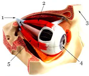
1. Optic nerve; 2. Muscle; 3. frontal bone; 4. Cornea; 5. Muscle
The front part of the eyeball - the cornea - is transparent, like glass: it transmits light into the eye. Then the light is captured by the "diaphragm" of the eye - the iris - and collected in a beam. Pigment cells of the iris give the eyes a certain color. If there is a lot of pigment, the eyes are colored in Brown color, if it is small or not at all - in greenish-gray and blue tones. The light then enters the pupil, a hole in the iris surrounded by two small muscles. In bright light, one muscle constricts the pupil, the other expands it, if it is dark. After passing the pupil, the light rays fall directly on the lens - an elastic organ that is constantly trying to take the shape of a ball. A ring of muscles interferes with it: they are constantly stretched and reduce the bulge of the lens. So, the lens easily changes its curvature. Therefore, the rays of light fall precisely on the layer of the retina dotted with rods and cones, and we clearly see objects. When we look at nearby objects, the lens becomes convex and refracts rays more strongly, and when objects that are far away from us, it becomes flatter and refracts rays more weakly. With age, the lens loses its elasticity. In order to somehow correct the trouble, we have to help our natural lens - the lens - and use glasses.
Like a camera, the eye is equipped with a "lens window", a "diaphragm", an "adjustable lens" and a "photosensitive layer" resembling photographic film. Only this layer is part of the eye itself, its retina. And yet a person sees more than a camera. After all, he looks at the world with two eyes. Both the left and right eyes see objects differently. Our brain compares the two received images and judges from them the form of what it sees. That is why people have spatial vision. But, for example, in a chicken, the eyes are set on the sides of the head, and it is not endowed with three-dimensional vision.
Nearsightedness and farsightedness
Almost one in three suffers from visual impairments. Nearsightedness and farsightedness are the most common, but are very well corrected with glasses or contact lenses. Myopia occurs as a result of pathology of the eye. myopic man can see clearly up close, but when looking at a distance, the image becomes very blurry. Farsightedness is a consequence of the normal aging of the eye. From the age of 40, we see less and less clearly up close, as the lens loses its flexibility over the years.
The eye is a complex and very delicate mechanism. His robot is still not fully understood by biologists. Although science is constantly trying to create something similar to the human eye. Sometimes it really does work. Now many people have some kind of device that is similar in function, operation and structure to the human eye - this is a camera and a video camera. What is similar between these devices and our eye? Now we'll find out.
The shape of the human eye resembles an irregular ball with a diameter of 2.5 cm and is called the eyeball in science. When we see something, light enters our eye. This light is nothing but reflections of what we are looking at. Light comes in the form of signals to back eyeball - retina. The retina is made up of many layers, but the main parts are rods and cones.
It is on the retina that the information that we have seen is processed and it is through it that the signal is transmitted to the brain. In order for the retina to be able to focus on the necessary object, there is a so-called lens in the eye. It is located in front of the eyeball and is natural in structure and shape. biconvex lens. The lens focuses information exactly on the necessary subject. In general, the lens is one of the most complex and "smart" parts of the eye. He owns accommodation - this is the ability to change his position, size and strength of refraction for better focusing. The lens changes its curvature depending on the situation - if we need to see nearby objects, the lens increases curvature, refracts light more and becomes convex. This helps to consider all the details to the smallest detail.
If we look at objects that are far away, the lens becomes flat and reduces its refractive power. All this he can do thanks to the ciliary muscle. But, of course, the lens itself cannot cope - it helps vitreous body.
This substance occupies 2/3 of the eyeball and consists of a jelly-like tissue. The vitreous body, in addition to refraction of light, also provides the eye with shape and incompressibility. Light enters the lens through the pupil. It can be seen in the mirror - this is the blackest circle in the central part of our eyes. The pupil can change its diameter and accordingly control the amount of incoming light. The muscles of the iris help him in this. We see it as a circle around the pupil, and as we know, this part of the eye can have different colors, it is precisely the pigment cells of the iris that determine this.
So, the pupil changes its size depending on the amount of light directed at it. If you look at your eyes in the mirror, you can see a lot of interesting things. If our eye looks at bright light, the pupil constricts and thus does not allow bright light to enter. in large numbers get on the retina.
If the environment is dark, the pupil dilates. Thus, this black circle does not destroy our vision. The sclera is located in front of the eye protein shell, 0.3-1 mm in diameter. This layer of the eyeball is made up of protein fibers and collagen cells. The sclera protects the eye and performs a supporting function. Its color is white with a certain milky tint, only in the central part it passes into the cornea - a transparent film.
The cornea is located above the pupil and iris and it is in it that light is refracted at the very beginning. Under the protein coat there is a choroid, where the pupil and iris are located. Thin blood capillaries also lie here, through which the eye receives the necessary substances from the blood.
Per vascular layer there is a ciliary body that contains the ciliary muscle, which means that light is bent in it. Between all these shells there are spaces, they are filled with a light-refracting transparent liquid that saturates the eye.
The outer parts of the eye are the eyelids - lower and upper. They contain the lacrimal glands, with the help of which the eyeball is moistened and protected from specks. There are muscles under the eyelids. There are only 3 pairs of them and all of them are engaged in the movement of the eye - some move the eye from left to right, others up and down, and others rotate it along the axis. These muscles pull the eye forward when looking at something up close and round it when looking far away.
Everything is very harmonious and absolutely all parts of the eye are involved in the process of focusing. If something is wrong with the optical apparatus, diseases such as myopia and hyperopia develop. With these vision diseases, light entering the eye does not fall on the retina, but on the area in front of it or behind it. With such changes in the optical system of the eye, near or distant objects become blurred.
Myopia is characterized by stretching of the sclera in the direction back and forth, and the eyeball takes the form of an ellipse. Through this, the axis was lengthened, and the light was focused not on the retina, but in front of it. A person with this disease wears lenses to reduce the refraction of light with a minus sign, since all distant objects are not at all clear. With farsightedness, on the contrary, all information falls behind the retina, and the apple itself is shortened along. With farsightedness, only glasses with a plus sign help well.
So, having examined all the main parts of the eye and understanding how they work, we can draw some conclusions - a light beam through eye cornea enters the retina, passing the vitreous body and the lens, enters the cones and rods, which process information.
What is interesting is that the image that hits the retina is not at all what you see. It is reduced in size and upside down. Why do we see the world correctly? Everything is done by our brain, when it receives information, it analyzes it and makes the necessary corrections and changes. But we begin to see everything, as it is necessary only in 3 weeks.
Babies up to this age see everything upside down, only then the brain begins to turn everything upside down as necessary. By the way, there have been many works and many experiments on this topic. So, for example, if a person puts on glasses that turn everything around, then at first the person is generally lost in space, but soon the brain normally perceives changes and new coordination skills are formed in it. Having taken off such glasses, the person again cannot understand what happened and again rebuilds his visual coordination and again sees everything correctly. Such possibilities of our visual apparatus and the visual center of the brain once again prove the flexibility and complexity of the structure of all systems of the human body.
The eyes are one of the main human tools for obtaining information about the world around us. From 80 to 90 percent of sensations people receive precisely through vision.
With the help of the eyes, a person recognizes the shape and color of objects, can track their movement in space. Without vision in modern world life is hard enough: a large proportion of incoming information is designed for visual perception. The device of the human eye allows it to be one of the most advanced optical instruments.
What do we see?
The function of vision in humans is carried out not only by the eyes - paired organ located in the eye sockets of the skull. Part visual analyzer also includes the optic nerve and whole system auxiliary systems: eyelids, lacrimal glands and muscles of the eyeball.
By the way, the latter are rightfully considered one of the fastest muscles in the human body. Even if the gaze is focused on one point, in one second these muscles allow the eyes to make more than a hundred synchronous movements.
Behind the eye, in the cavity of the orbit, there is a kind of "buffer" of adipose tissue, and the closed parts of the eyeball are protected by the conjunctiva - the mucous membrane of the eye, penetrated by blood vessels.
The eyeball in all people is about the same size. Since birth, it has approximately doubled in size.
How do we see?
The human eye is a complex optical system, consisting of several lenses and a special sensor that perceives the image.
First, light rays enter the pupil located behind the cornea of the eye, which is the first lens of the system.
The pupil is analogous to the diaphragm in a camera. It is located in the center of the iris and is able to narrow and expand, adjusting the intensity of the light flux entering the eye.
The pupil is able to pass only those light rays that are located directly in front of it, and the pigment of the iris delays side rays that can cause image distortion.
lens
The light rays passing through the pupil are refracted by the lens - the second lens of the eye. The shape of the lens can be changed with the help of a special muscle.
To focus on closer objects, the muscle tenses and the lens becomes more convex. If focusing on distant objects is required, the muscle relaxes and the lens becomes flat. This process is called accommodation.
If it is disturbed, due to the weakness of the muscles of the lens, it develops myopia(inability to distinguish distant objects) and farsightedness(difficulty distinguishing closely spaced objects)
Behind the lens is the vitreous body. It occupies almost the entire cavity of the eye up to the retina itself and provides the elasticity of the eyeball.
Receiving device - retina
After focusing with the lens, the rays of light fall on the retina - a kind of concave screen, on which an inverted image of what is seen is projected.
The outer layer of the retina consists of two types of special cells: rods that perceive light and cones that recognize colors. With help chemical processes stimulation of these cells by light is encoded into a nerve impulse, which is transmitted to the brain.
The most sensitive part of the retina, which allows you to distinguish colors and fine details of objects - yellow spot or macula, located in its center.
There is also a blind spot on the retina - an area completely devoid of rods and cones. Here, the optic nerve emerges from the retina, which transmits the encoded image to the brain, where it is finally processed and interpreted.
eye diseases
There are many eye diseases. Some of them are caused by disorders in the eye itself, the rest affect the eyes when common diseases and consequences wrong image life: at diabetes, problems with gland functions internal secretion, hypertension, alcohol consumption and so on.
The eyes are one of the main human tools for obtaining information about the world around us. This paired organ is a complex system of two lenses and a receiving device - the retina.
Visual impairment is one of the consequences of an unhealthy lifestyle.
The human eye is often cited as an example of amazing natural engineering - but judging by the fact that this is one of the 40 devices that appeared during the evolution of different organisms, we should moderate our anthropocentrism and admit that the structure of the human eye is not something perfect.
The story about the eye is best to start with a photon. A quantum of electromagnetic radiation slowly flies strictly into the eye of an unsuspecting passer-by, who squints from an unexpected glare from someone's watch.
The first part of the optical system of the eye is the cornea. It changes the direction of the light. This is possible due to such a property of light as refraction, which is also responsible for the rainbow. The speed of light is constant in a vacuum - 300,000,000 m/s. But when moving from one medium to another (in this case, from air to the eye), light changes its speed and direction of movement. For air, the refractive index is 1.000293, for the cornea - 1.376. This means that the light beam in the cornea slows down its movement by 1.376 times and deviates closer to the center of the eye.
A favorite way to split partisans is to shine a bright lamp in their face. It hurts for two reasons. Bright light- this is a powerful electromagnetic radiation: trillions of photons attack the retina, and its nerve endings are forced to transmit a frantic amount of signals to the brain. From overvoltage, nerves, like wires, burn out. The muscles in the iris are forced to contract as hard as they can, in a desperate attempt to close the pupil and protect the retina.
And flies up to the pupil. Everything is simple with him - this is a hole in the iris. Due to the circular and radial muscles, the iris can constrict and expand the pupil accordingly, regulating the amount of light entering the eye, like a diaphragm in a camera. Human pupil diameter can vary from 1 to 8 mm depending on the illumination.
Having flown through the pupil, the photon hits the lens - the second lens responsible for its trajectory. The lens refracts light less than the cornea, but it is mobile. The lens hangs on cylindrical muscles that change its curvature, thereby allowing us to focus on objects at different distances from us.
It is with the focus that visual impairments are associated. The most common are nearsightedness and farsightedness. The image in both cases does not focus on the retina, as it should, but in front of it (nearsightedness), or behind it (farsightedness). The eye is to blame for this, which changes shape from round to oval, and then the retina moves away from the lens or approaches it.
After the lens, the photon flies through the vitreous body (transparent jelly - 2/3 of the volume of the entire eye, 99% - water) straight to the retina. This is where photons are registered and arrival messages are sent along the nerves to the brain.
The retina is lined with photoreceptor cells: when there is no light, they produce special substances - neurotransmitters, but as soon as a photon enters them, photoreceptor cells stop producing them - and this is a signal to the brain. There are two types of these cells: rods, which are more sensitive to light, and cones, which are better at detecting movement. We have about a hundred million rods and another 6-7 million cones, in total more than a hundred million light-sensitive elements - this is more than 100 megapixels, which no "Hassel" could dream of.
A blind spot is a breakthrough point where there are no light-sensitive cells at all. It is quite large - 1-2 mm in diameter. Fortunately we have binocular vision and there is a brain that combines two pictures with spots into one normal one.
At the time of signal transmission human eye there is a problem with logic. The underwater octopus, which does not really need vision, is much more consistent in this sense. In octopuses, a photon first hits a layer of cones and rods on the retina, just behind which a layer of neurons waits and transmits a signal to the brain. In humans, light first breaks through the layers of neurons - and only then hits the photoreceptors. Because of this, there is a first spot in the eye - a blind spot.
The second spot is yellow, this is the central area of the retina directly opposite the pupil, slightly higher optic nerve. This place sees the eye best: the concentration of light-sensitive cells here is greatly increased, so our vision in the center of the visual field is much sharper than peripheral.
The image on the retina is inverted. The brain knows how to correctly interpret the picture, and restores the original image from the inverted one. Children see everything upside down for the first couple of days while their brain sets up its photoshop. If you put on glasses that flip the image (this was first done back in 1896), then in a couple of days our brain will learn to interpret such an inverted picture correctly.