Epithelial tissues perform the following functions. Epithelial tissues general information
Tissue is a collection of cells and intercellular substance. She has general signs structures and performs the same functions. There are four types of tissues in the body: epithelial, nervous, muscle and connective.
The structure of epithelial and animal tissue is determined, first of all, by its localization. Epithelial tissue is the boundary layer of cells lining the body, mucous membranes internal organs and cavities. Also, many glands in the body are formed by epithelium.
general characteristics
The structure of epithelial tissue has a number of features inherent only to epithelium. main feature lies in the fact that the tissue itself looks like a continuous layer of cells that fit tightly together.
The epithelium lining all surfaces in the body has the appearance of a layer, while in the liver, pancreas, thyroid, salivary and other glands it is a cluster of cells. In the first case, it is located on top of the basement membrane, which separates the epithelium from the connective tissue. But there are exceptions when the structure of epithelial and connective tissue is considered in the context of their interaction. In particular, in the lymphatic system there is an alternation of epithelial and connective tissue cells. This type epithelium is called atypical.
High regenerative capacity is another feature of the epithelium.
The cells of this tissue are polar, which is due to the difference in the basal and apical parts of the cell center.
The structure of epithelial tissue is largely explained by its border position, which, in turn, makes the epithelium an important link in metabolic processes. This tissue is involved in the absorption of nutrients from the intestines into the blood and lymph, in the excretion of urine through the epithelium of the kidneys, etc. We must also not forget about the protective function, which is to protect tissues from damaging influences.
The structure of the substance that forms the basement membrane shows what it contains a large number of mucopolysaccharides, and also has a network of thin fibrils.
How is epithelial tissue formed?
The structural features of epithelial tissue in animals and humans are largely dictated by the fact that its development is carried out from all three This feature inherent only to this type of fabric. The ectoderm gives rise to the epithelium of the skin, oral cavity, a significant part of the esophagus, and the cornea of the eye; endoderm - epithelium gastrointestinal tract; and mesoderm - epithelium genitourinary organs and serous membranes.
IN embryonic development begins to form at the very early stages. Since it is part of the placenta sufficient quantity epithelial tissue, it is a participant in the metabolism between the mother and the embryo.
Maintaining the integrity of epithelial cells
The interaction of neighboring cells in the layer is possible due to the presence of desmosomes. These are special multiple structures of submicroscopic size that consist of two halves. Each of them, thickening in certain places, occupies adjacent surfaces of neighboring cells. In the slit-like space between the halves of the desmosomes there is a substance of carbohydrate origin.
In cases where the intercellular spaces are wide, desmosomes are located at the ends of cytoplasmic protrusions directed towards each other on the contacting cells. If you examine a pair of these protrusions under a microscope, you will find that they have the appearance of an intercellular bridge.
IN small intestine the integrity of the layer is maintained due to the fusion of the cell membranes of neighboring cells at the points of contact. Such places are often called end plates.
There are other cases where there are no special structures to ensure integrity. Then the contact of neighboring cells occurs due to the contact of smooth or curved cell surfaces. The edges of the cells may overlap each other in a tiled manner.
The structure of an epithelial tissue cell
Features of epithelial tissue cells include the presence of a plasma membrane on their surface.
In cells involved in the release of metabolic products, folding is observed in the plasma membrane of the basal part of the cell body.
Epithelial cells are the scientific name for cells that form epithelial tissue. The structural features and functions of epithelial cells are closely interrelated. So, according to their shape, they are divided into flat, cubic and columnar. Euchromatin predominates in the nucleus, due to which it has a light color. The nucleus is quite large, its shape coincides with the shape of the cell.
The pronounced polarity determines the location of the nucleus in the basal part, above it there are mitochondria, the Golgi complex and centrioles. In cells performing a secretory function, the endoplasmic reticulum and Golgi complex are especially well developed. The epithelium, which experiences a large mechanical load, has a system of special threads in its cells - tonofibrils, which create a kind of barrier designed to protect the cells from deformation.
Microvilli
Some cells, or rather their cytoplasm, can form tiny ones on the surface, directed towards outside, outgrowths - microvilli. Their largest accumulations are present on the apical surface of the epithelium in small intestine and the main sections of the convoluted tubules of the kidneys. Due to the parallel arrangement of microvilli in the cuticles of the intestinal epithelium and the brush border of the kidneys, stripes are formed that can be viewed under an optical microscope. In addition, microvilli in these places contain a number of enzymes.
Classification
Features of the structure of epithelial tissues different localization allow them to be classified according to several criteria.
Depending on the shape of the cells, the epithelium can be cylindrical, cubic and flat, and depending on the location of the cells - single-layer and multilayer.

Glandular epithelium is also isolated, which performs a secretory function in the body.

Single layer epithelium
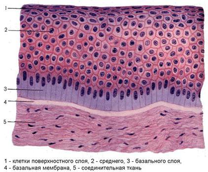
The name of single-layer epithelium speaks for itself: in it, all cells are located on the basement membrane in one layer. If the shape of all cells is the same (that is, they are isomorphic) and are at the same level, then they speak of single-row epithelium. And if in single layer epithelium alternation of cells is observed various shapes, their nuclei are located at different levels, then this is a multirow or anisomorphic epithelium.

Stratified epithelium
In stratified epithelium, only the bottom layer is in contact with the basement membrane, and the other layers are above it. Cells of different layers differ in shape. The structure of epithelial tissue of this type allows us to distinguish several types of multilayered epithelium depending on the shape and condition: stratified squamous, multilayered keratinized (there are keratinized scales on the surface), multilayered non-keratinized.

There is also the so-called transitional epithelium lining the organs excretory system. Depending on whether or not it is stretched, the fabric acquires different type. Thus, when the bladder is stretched, the epithelium is in a thinned state and forms two layers of cells - basal and integumentary. And when bladder is in a compressed (shortened) form, the epithelial tissue thickens sharply, the cells of the basal layer become polymorphic and their nuclei are at different levels. The integumentary cells acquire a pear-shaped shape and are layered on top of each other.
Histogenetic classification of epithelia
The structure of epithelial tissue in animals and humans often becomes the subject of scientific and medical research. In these cases, the histogenetic classification developed by Academician N. G. Khlopin is used more often than others. According to it, there are five types of epithelium. The criterion is from which rudiments the tissue developed during embryogenesis.
1. Epidermal type, which originated from the ectoderm and prechordal plate.
2. Enterodermal type, the development of which originated from the intestinal endoderm.
3. Coelonephrodermal type, developed from the coelomic lining and nephrotome.
4. Angiodermal type, the development of which began from the area of mesenchyme that forms vascular endothelium, which is called angioblast.
5. Ependymoglial type, which originated from the neural tube.
Features of the structure of epithelial tissues forming glands
The glandular epithelium performs a secretory function. This type of tissue is a collection of glandular (secretory) cells called granulocytes. Their function is to carry out synthesis, as well as the release of specific substances - secrets.
It is thanks to secretion that the body is able to perform many important functions. The glands secrete secretions on the surface of the skin and mucous membranes, inside the cavities of a number of internal organs, as well as into the blood and lymph. In the first case, we are talking about exocrine, and in the second, about endocrine secretion.

Exocrine secretion allows milk production (in female body), gastric and intestinal juice, saliva, bile, sweat and sebum. Secrets endocrine glands are hormones that perform humoral regulation in organism.
The structure of epithelial tissue of this type may be different due to the fact that granulocytes can take different shapes. It depends on the phase of secretion.
Both types of glands (endocrine and exocrine) can consist of a single cell (unicellular) or many cells (multicellular).
Characteristic morphological features of epithelial tissues
Epithelial tissues are a collection of polarly differentiated cells, closely adjacent to each other, located in the form of a layer on the basement membrane; they are missing blood vessels and very little or no intercellular substance.
Functions. Epithelia cover the surface of the body, secondary body cavities, internal and outer surface hollow internal organs, form secretory sections and excretory ducts exocrine glands. Their main functions: delimiting, protective, suction, secretory, excretory.
Histogenesis. Epithelial tissues develop from all three germ layers. Epithelia of ectodermal origin are predominantly multilayered, while those developing from endoderm are always single-layered. Both single-layer and multilayer epithelia develop from the mesoderm.
Classification of epithelial tissues
1. Morphofunctional classification takes into account the structural features and functions performed by one or another type of epithelium.
Based on their structure, epithelia are divided into single-layer and multilayer. Main principle this classification is the ratio of cells to the basement membrane (Table 1). The functional specificity of single-layer epithelia is usually determined by the presence of specialized organelles. For example, in the stomach the epithelium is single-layered, prismatic, single-row glandular. The first three definitions characterize structural features, and the last indicates that gastric epithelial cells perform a secretory function. In the intestine, the epithelium is single-layered, prismatic, single-row, bordered. The presence of a brush border in epithelial cells suggests an absorptive function. In the airways, in particular in the trachea, the epithelium is single-layered, prismatic, multirow ciliated (or ciliated). It is known that cilia play a role in this case protective function. Multilayer epithelia perform protective and glandular functions.
Table 1. Comparative characteristics single-layer and multilayer epithelium.
|
SINGLE LAYER EPITHELIAS |
MULTILAYERED EPITHELIA |
|
All epithelial cells are in contact with the basement membrane: |
Not all epithelial cells are in contact with the basement membrane: |
|
1) single-layer flat; 2) single-layer cubic (low prismatic); 3) single-layer prismatic (cylindrical, columnar) Happens: |
1) multilayer flat non-keratinizing contains three layers of different cells: basal, intermediate (spinous) and superficial; 5 layers: basal, spinous, granular, shiny and horny; The basal and spinous layers constitute the germinal layer of the epithelium, since the cells of these layers are capable of division. |
Ontophylogenetic classification (according to N. G. Khlopin). This classification takes into account from which embryonic rudiment a particular epithelium developed. According to this classification, epidermal (skin), enterodermal (intestinal), coelonephrodermal, ependymoglial and angiodermal types of epithelium are distinguished.
For example, cutaneous epithelium covers the skin, lining oral cavity, esophagus, glandless chambers of the multichamber stomach, vagina, urethra, border section of the anal canal; intestinal-type epithelium lines the single-chamber stomach, abomasum, and intestines; epithelium of the coelonephrodermal type lines the body cavities (mesothelium of the serous membranes), forms renal tubules; ependymoglial type of epithelium lines the ventricles of the brain and the central canal spinal cord; angiodermal epithelium lines the cavities of the heart and blood vessels.
Single-layer and multilayer epithelia are characterized by the presence of special organelles - desmosomes, hemidesmosomes, tonofilaments and tonofibrils. In addition, single-layer epithelia may have cilia and microvilli on the free surface of cells (see section “Cytology”).
All types of epithelia are located on the basement membrane (Fig. 7). The basement membrane consists of fibrillar structures and an amorphous matrix containing complex proteins - glycoproteins, proteoglycans and polysaccharides (glycosaminoglycans).
Rice. 7. Scheme of the structure of the basement membrane (according to Yu. K. Kotovsky).
BM – basement membrane; WITH - Light plate; T – dark plate. 1 – cytoplasm of epithelial cells; 2 – core; 3 – hemidesmosomes; 4 – keratin tonofilaments; 5 – anchor filaments; 6 – plasmalemma of epithelial cells; 7 – anchoring filaments; 8 – loose connective tissue; 9 – Hemocapillary.
The basement membrane regulates the permeability of substances (barrier and trophic function) and prevents invasion of the epithelium into the connective tissue. The glycoproteins it contains (fibronectin and laminin) promote the adhesion of epithelial cells to the membrane and induce their proliferation and differentiation during the regeneration process.
By location and function of the epithelium are divided into: superficial (cover organs from the outside and inside) and glandular (form the secretory sections and excretory ducts of the exocrine glands).
Surface epithelia are border tissues that separate the body from the external environment and participate in the exchange of substances and energy between the body and the external environment environment. They are located on the surface of the body (integumentary), the mucous membranes of internal organs (stomach, intestines, lungs, heart, etc.) and secondary cavities (lining).
Glandular epithelia have pronounced secretory activity. Glandular cells - glandulocytes are characterized by a polar arrangement of organelles general meaning, well-developed ER and Golgi complex, the presence of secretory granules in the cytoplasm.
The process of functional activity of a glandular cell associated with the formation, accumulation and release of secretions beyond its boundaries, as well as restoration of the cell after secretion is released, is called Secretory cycle.
During the secretory cycle, initial products (water, various inorganic substances and low molecular weight organic compounds: amino acids, monosaccharides, fatty acid etc.), from which, with the participation of organelles of general importance, a secret is synthesized and accumulated in cells, and then, through exocytosis, is released into the external ( Exocrine glands ) or internal ( Endocrine glands ) Wednesday.
Secretion is released (extrusion) by diffusion or in the form of granules, but can also be by converting the entire cell into a common secretory mass.
Regulation of the secretory cycle is carried out with the participation of humoral and nervous mechanisms.
Epithelial regeneration
For various types epithelium is characterized by high regenerative activity. It is carried out due to the cambial elements, which divide by mitosis, constantly replenishing the loss of worn-out cells. Glandular cells, which secrete according to the merocrine and apocrine type, are, in addition, capable of maintaining their vital functions not only through reproduction, but also due to intracellular regeneration. In holocrine glands, constantly dying glandulocytes are replaced during the secretory cycle due to the division of stem cells located on the basement membrane (cellular regeneration).
Types of epithelium
- Single layer squamous epithelium(endothelium and mesothelium). The endothelium lines the inside of blood vessels, lymphatic vessels, cavities of the heart. Endothelial cells are flat, poor in organelles and form the endothelial layer. The metabolic function is well developed. They create conditions for blood flow. When the epithelium is damaged, blood clots form. The endothelium develops from mesenchyme. The second type - mesothelium - develops from mesoderm. Lines all serous membranes. Consists of flat polygonal shape cells connected to each other by uneven edges. Cells have one, rarely two, flattened nuclei. There are short microvilli on the apical surface. They have absorptive, excretory and delimiting functions. The mesothelium ensures the free sliding of internal organs relative to each other. The mesothelium secretes a mucous secretion onto its surface. The mesothelium prevents the formation of connective tissue adhesions. They regenerate quite well due to mitosis.
- Single layer cuboidal epithelium develops from endoderm and mesoderm. On the apical surface there are microvilli that increase the working surface, and in the basal part the cytolemma forms deep folds, between which mitochondria are located in the cytoplasm, so the basal part of the cells looks striated. Lines the small excretory ducts of the pancreas, bile ducts and renal tubules.
- Single layer columnar epithelium found in the organs of the middle section of the digestive canal, digestive glands, kidneys, gonads and genital tract. In this case, the structure and function are determined by its localization. Develops from endoderm and mesoderm. The gastric mucosa is lined with single-layer glandular epithelium. It produces and secretes a mucous secretion that spreads over the surface of the epithelium and protects the mucous membrane from damage. The cytolemma of the basal part also has small folds. The epithelium has high regeneration.
- The kidney tubules and intestinal mucosa are lined bordered epithelium. In the bordered epithelium of the intestine, border cells - enterocytes - predominate. At their top there are numerous microvilli. In this zone, parietal digestion and intensive absorption of food occur. Mucous goblet cells produce mucus on the surface of the epithelium, and small endocrine cells are located between the cells. They secrete hormones that provide local regulation.
- Single layer multirow ciliated epithelium. It lines the airways and is of ectodermal origin. In it, cells are of different heights, and the nuclei are located at different levels. The cells are arranged in a layer. Under the basement membrane lies loose connective tissue with blood vessels, and the epithelial layer is dominated by highly differentiated ciliated cells. They have a narrow base and a wide top. At the top there are flickering cilia. They are completely immersed in mucus. Between the ciliated cells are goblet cells - these are single-celled mucous glands. They produce a mucous secretion onto the surface of the epithelium.
There are endocrine cells. Between them there are short and long intercalary cells, these are stem cells, poorly differentiated, due to them cell proliferation occurs. The ciliated cilia perform oscillatory movements and move the mucous film along the airways to the external environment.
- Stratified squamous non-keratinizing epithelium. It develops from the ectoderm, lining the cornea, anterior section the digestive canal and the anal section of the digestive canal, vagina. The cells are arranged in several layers. On the basement membrane lies a layer of basal or columnar cells. Some of them are stem cells. They proliferate, separate from the basement membrane, transform into polygonal cells with projections, spines, and the combination of these cells forms a layer of spinous cells arranged in several floors. They gradually flatten and form surface layer flat, which are torn off from the surface in external environment.
- Stratified squamous keratinizing epithelium- epidermis, it lines skin. In thick skin (palm surfaces), which is constantly under stress, the epidermis contains 5 layers:
- 1 – basal layer – contains stem cells, differentiated cylindrical and pigment cells (pigmentocytes).
- 2 – stratum spinosum – polygonal cells containing tonofibrils.
- 3 – granular layer – the cells acquire a rhomboid shape, the tonofibrils disintegrate and inside these cells the protein keratohyalin is formed in the form of grains, this is where the process of keratinization begins.
- 4 – stratum lucidum – a narrow layer, in which the cells become flat, they gradually lose their intracellular structure, and keratohyalin turns into eleidin.
- 5 – stratum corneum – contains horny scales that have completely lost their cell structure and contain the protein keratin. With mechanical stress and deterioration of blood supply, the process of keratinization intensifies.
Thin skin that does not experience stress lacks a grainy and shiny layer.
- Multilayer cubic and columnar epithelium are extremely rare - in the area of the conjunctiva of the eye and the area of \u200b\u200bthe junction of the rectum between single-layer and multilayer epithelium.
- Transitional epithelium(uroepithelium) lines urinary tract and allantois. Contains a basal layer of cells, some of the cells gradually separate from the basement membrane and form an intermediate layer of pyriform cells. On the surface there is a layer of integumentary cells - large cells, sometimes double-rowed, covered with mucus. The thickness of this epithelium varies depending on the degree of stretching of the wall urinary organs. The epithelium is capable of secreting a secretion that protects its cells from the effects of urine.
- Glandular epithelium- a type of epithelial tissue that consists of epithelial glandular cells, which in the process of evolution acquired the leading property of producing and secreting secretions. Such cells are called secretory (glandular) - glandulocytes. They have exactly the same general characteristics as covering epithelium. Among the epithelial cells there are secretory cells, of which there are 2 types.
- exocrine - release their secretion into the external environment or the lumen of the organ.
- endocrine - release their secretions directly into the bloodstream.
Located in the glands of the skin, intestines, salivary glands, glands internal secretion and etc.
Characteristics
Main features epithelial tissues - fast regeneration and lack of blood vessels.
Classification.
There are several classifications of epithelia, which are based on various signs: origin, structure, functions. Of them greatest distribution received a morphological classification that takes into account mainly the relationship of cells to the basement membrane and their shape.
Single layer epithelium can be single-row or multi-row. In single-row epithelium, all cells have the same shape - flat, cubic or prismatic, their nuclei lie at the same level, i.e. in one row. Such epithelium is also called isomorphic.
Stratified epithelium It can be keratinizing, non-keratinizing and transitional. The epithelium in which keratinization processes occur, associated with the differentiation of cells of the upper layers into flat horny scales, is called multilayered squamous keratinization. In the absence of keratinization, the epithelium is called stratified squamous non-keratinizing.
Transitional epithelium lines organs subject to strong stretching - the bladder, ureters, etc. When the volume of an organ changes, the thickness and structure of the epithelium also changes.
Along with morphological classification, it is used ontophylogenetic classification, created by Russian histologist N. G. Khlopin. It is based on the peculiarities of the development of epithelia from tissue primordia.
Epidermal type The epithelium is formed from the ectoderm, has a multilayer or multirow structure, and is adapted to perform primarily a protective function.
Enterodermal type The epithelium develops from the endoderm, is single-layer prismatic in structure, carries out the processes of absorption of substances, and performs a glandular function.
Coelonephrodermal type epithelium develops from mesoderm, single-layer, flat, cubic or prismatic in structure; performs a barrier or excretory function.
Ependymoglial type represented by a special epithelium lining, for example, the cavities of the brain. The source of its formation is the neural tube.
see also
Wikimedia Foundation. 2010.
See what “Epithelial tissue” is in other dictionaries:
epithelial tissue- Rice. 1. Single-layer epithelia. Rice. 1. Single-layer epithelia: A prismatic bordered; B multirow prismatic ciliated; B cubic; G flat; 1 prismatic cells; 2 connective tissue; ... Veterinary encyclopedic Dictionary
- (epithelium), a layer of closely spaced cells covering the surface of the body and lining all its cavities. Most glands also consist of epithelium (glandular epithelium). Flat epithelium consists of flattened cells that have the shape... ... Biological encyclopedic dictionary
epithelial tissue- dermis shell. hypodermis. endoderm. epithelium. endothelium. mesothelium. ependyma. sarcolemma. epicardium pericardium. endocardium sclera. hymen. pleura...
This term has other meanings, see Fabric (meanings). Tissue is a system of cells and intercellular substance, united by a common origin, structure and functions. Science studies the structure of tissues of living organisms... ... Wikipedia
animal tissue- fabrics: connective. epithelial. muscular. nervous. body. flesh. meat muscle(vomited a piece of meat). pulp. histogenesis. blastema. mesoglea. slime. slimy. transudate transudation. exudate exudation. tissue fluid... Ideographic Dictionary of the Russian Language
A historically established community of cells and intercellular substance, united by a unity of origin, structure and function. There are four types of tissues in the human body: epithelial, connective, muscle and nervous. Each fabric... Medical terms - Buraya adipose tissue... Wikipedia
Epithelium(Latin epithelium, from other Greek - nipple of the mammary gland), or epithelial tissue- a layer of cells lining the surface (epidermis) and body cavities, as well as the mucous membranes of internal organs, the digestive tract, respiratory system, genitourinary tract. In addition, it forms most of the glands of the body.
Morphological classification of epithelium:
- Single layer epithelium can be single-row or multi-row. U single-row single-layer epithelium all cells have the same shape - flat, cubic or prismatic, their nuclei lie at the same level, that is, in one row. In multirow single-layer epithelium, hematoxylin-eosin-stained, prismatic and intercalary cells are distinguished; the latter, in turn, are divided according to the principle of the ratio of the nucleus to the basement membrane into high intercalary and low intercalary cells.
- Stratified epithelium It can be keratinizing, non-keratinizing and transitional. The epithelium in which keratinization processes occur, associated with the differentiation of cells of the upper layers into flat horny scales, is called multilayered squamous keratinizing epithelium. In the absence of keratinization, the epithelium is called stratified squamous non-keratinizing epithelium.
- Transitional epithelium lines organs subject to strong stretching - the bladder, ureters, etc. When the volume of an organ changes, the thickness and structure of the epithelium also changes.
Ontophylogenetic classification of epithelium:
Along with the morphological classification of the epithelium, the ontophylogenetic classification of the epithelium, created by the Russian histologist N. G. Khlopin, is used. The ontophylogenetic classification of epithelium is based on the features of the development of epithelia from tissue primordia.
- Epidermal type of epithelium It is formed from the ectoderm, has a multilayer or multirow structure, and is adapted to perform primarily a protective function.
- Endodermal type of epithelium develops from the endoderm, is single-layered prismatic in structure, carries out the processes of absorption of substances, and performs a glandular function.
- Coelonephrodermal type of epithelium develops from the mesoderm, single-layer, flat, cubic or prismatic in structure; performs a barrier or excretory function.
- Ependymoglial type of epithelium represented by a special epithelium lining, for example, the cavities of the brain. The source of epithelial formation is the neural tube.
- Angiodermal type of epithelium formed from mesenchyme, lining blood vessels from the inside.
Types of epithelium
Single layer epithelium
- Single layer squamous epithelium(endothelium and mesothelium). The endothelium lines the inside of blood vessels, lymphatic vessels, and the cavities of the heart. Endothelial cells are flat, poor in organelles and form the endothelial layer. The metabolic function is well developed. They create conditions for blood flow. When the epithelium is damaged, blood clots form. The endothelium develops from mesenchyme. The second type - mesothelium - develops from mesoderm. Lines all serous membranes. Consists of flat polygonal cells connected to each other by uneven edges. Cells have one, rarely two, flattened nuclei. There are short microvilli on the apical surface. They have absorptive, excretory and delimiting functions. The mesothelium ensures the free sliding of internal organs relative to each other. The mesothelium secretes a mucous secretion onto its surface. The mesothelium prevents the formation of connective tissue adhesions. They regenerate quite well due to mitosis.
- Single layer cuboidal epithelium develops from endoderm and mesoderm. On the apical surface there are microvilli that increase the working surface, and in the basal part the cytolemma forms deep folds, between which mitochondria are located in the cytoplasm, so the basal part of the cells looks striated. Lines the small excretory ducts of the pancreas, bile ducts and renal tubules.
- Single layer columnar epithelium found in the organs of the middle part of the digestive canal, digestive glands, kidneys, gonads and genital tract. In this case, the structure and function are determined by its localization. Develops from endoderm and mesoderm. The gastric mucosa is lined with single-layer glandular epithelium. It produces and secretes a mucous secretion that spreads over the surface of the epithelium and protects the mucous membrane from damage. The cytolemma of the basal part also has small folds. Single-layer columnar epithelium has high regeneration.
- The kidney tubules and intestinal mucosa are lined bordered epithelium. In the bordered epithelium of the intestine, border cells - enterocytes - predominate. At their top there are numerous microvilli. In this zone, parietal digestion and intensive absorption of food occur. Mucous goblet cells produce mucus on the surface of the epithelium, and small endocrine cells are located between the cells. They secrete hormones that provide local regulation.
- Single layer multirow ciliated epithelium. It lines the airways and is of endodermal origin. In it, cells are of different heights, and the nuclei are located at different levels. The cells are arranged in a layer. Under the basement membrane lies loose connective tissue with blood vessels, and the epithelial layer is dominated by highly differentiated ciliated cells. They have a narrow base and a wide top. At the top there are flickering cilia. They are completely immersed in mucus. Between the ciliated cells are goblet cells - these are single-celled mucous glands. They produce a mucous secretion onto the surface of the epithelium. There are endocrine cells. Between them there are short and long intercalary cells, these are stem cells, poorly differentiated, due to them cell proliferation occurs. The ciliated cilia perform oscillatory movements and move the mucous film along the airways to the external environment.
Stratified epithelium
Multilayered squamous non-keratinizing epithelium.
Multilayered flat non-keratinizing epithelium develops from the ectoderm and lines the cornea, the anterior part of the digestive canal and the anal part of the digestive canal, and the vagina. The cells are arranged in several layers. On the basement membrane lies a layer of basal or columnar cells. Some of them are stem cells. Stem cells proliferate, separate from the basement membrane, transform into polygonal cells with projections, spines, and the combination of these cells forms a layer of spinous cells arranged in several floors. They gradually flatten and form a surface layer of flat ones, which are rejected from the surface into the external environment.
Stratified squamous keratinizing epithelium- epidermis, it lines the skin. In thick skin (palm surfaces), which is constantly under stress, the epidermis contains 5 layers:
- basal layer - contains stem cells, differentiated cylindrical and pigment cells (pigmentocytes).
- stratum spinosum - polygonal cells containing tonofibrils.
- granular layer - the cells acquire a rhomboid shape, the tonofibrils disintegrate and inside these cells the protein keratohyalin is formed in the form of grains, this is where the process of keratinization begins.
- The stratum lucidum is a narrow layer, in which the cells become flat, they gradually lose their intracellular structure, and keratohyalin turns into eleidin.
- stratum corneum - contains horny scales that have completely lost their cell structure and contain the protein keratin. With mechanical stress and deterioration of blood supply, the process of keratinization intensifies.
In thin skin that does not experience stress, there is no granular and shiny layer. Multilayer cubic and cylindrical epithelium are extremely rare - in the area of the conjunctiva of the eye and the area of the junction of the rectum between single-layer and multilayer epithelium.
Transitional epithelium (uroepithelium) lines the urinary tract and allantois. Contains a basal layer of cells, some of the cells gradually separate from the basement membrane and form an intermediate layer of pyriform cells. On the surface there is a layer of integumentary cells - large cells, sometimes double-rowed, covered with mucus. The thickness of this epithelium varies depending on the degree of stretching of the wall of the urinary organs. The epithelium is capable of secreting a secretion that protects its cells from the effects of urine.
Glandular epithelium- a type of epithelial tissue, which consists of epithelial glandular cells, which in the process of evolution acquired the leading property of producing and secreting secretions. Such cells are called secretory (glandular) - glandulocytes. They have exactly the same general characteristics as the integumentary epithelium. Located in the glands of the skin, intestines, salivary glands, endocrine glands, etc. Among the epithelial cells are secretory cells, of which there are 2 types:
- exocrine - release their secretion into the external environment or the lumen of an organ;
- endocrine - release their secretions directly into the bloodstream.
Characteristic features of the epithelium
There are five main characteristic features of the epithelium:
Epithelia are layers (less often strands) of cells - epithelial cells. There is almost no intercellular substance between them, and the cells are closely connected to each other through various contacts.
Epithelia are located on basement membranes that separate epithelial cells from the underlying connective tissue.
The epithelium has polarity. The two cell sections - basal (lying at the base) and apical (apical) - have different structures.
The epithelium does not contain blood vessels. Epithelial cells are nourished diffusely through the basement membrane from the side of the underlying connective tissue.
Epithelia have a high ability to regenerate. Epithelial restoration occurs due to mitotic division and differentiation of stem cells.
The epithelium is the layers covering the internal and external surfaces organisms. Its main function is to protect the relevant organs from mechanical damage and infections. In those places where body tissue is subjected to constant stress and friction and “wears out,” epithelial cells multiply at high speed. Often, in areas of high stress, the epithelium becomes denser or keratinized. The free surface of the epithelium can also perform the functions of absorption, secretion and excretion, and perceive irritations.
Epithelial cells held together by a cementitious substance containing hyaluronic acid. Since the epithelium does not have blood vessels, oxygen supply and nutrients occurs by diffusion through the lymphatic system. Nerve endings can penetrate the epithelium.
Depending on the shape of the cell and the number of cell layers, the epithelium is divided into several types.
The least specialized of all is cuboidal epithelium. Its cells, as the name suggests, are cubic in cross section. This type of epithelium lines the ducts of many glands and also performs secretory functions inside them.
Cells squamous epithelium thin and flattened; they are tightly connected to each other by protoplasmic bonds. Thanks to this, they do not interfere with diffusion various substances into the organs that these cells line: lung alveoli, capillary walls.
Tall and rather narrow cells columnar epithelium line the stomach and intestines. Scattered among the cylindrical cells, goblet cells secrete mucus that protects these organs from self-digestion, and at the same time create a lubricant that helps in the movement of food. Microvilli are often found on the free surface of cells, increasing the absorption surface.